Review
. 2017 Jun 20:86:845-872.
doi: 10.1146/annurev-biochem-101910-144233. Epub 2017 Mar 9.

Microbial Rhodopsins: Diversity, Mechanisms, and Optogenetic Applications

Affiliations
Review

Microbial Rhodopsins: Diversity, Mechanisms, and Optogenetic Applications

Elena G Govorunova et al. Annu Rev Biochem. .

Abstract

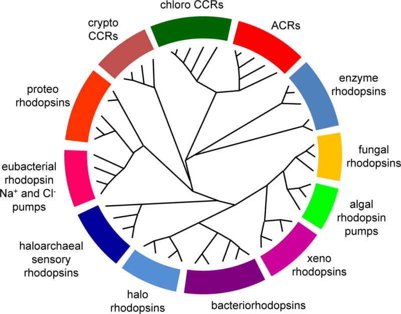
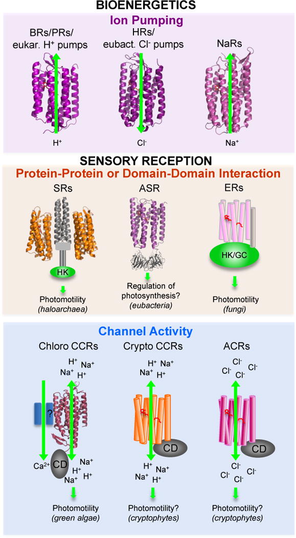
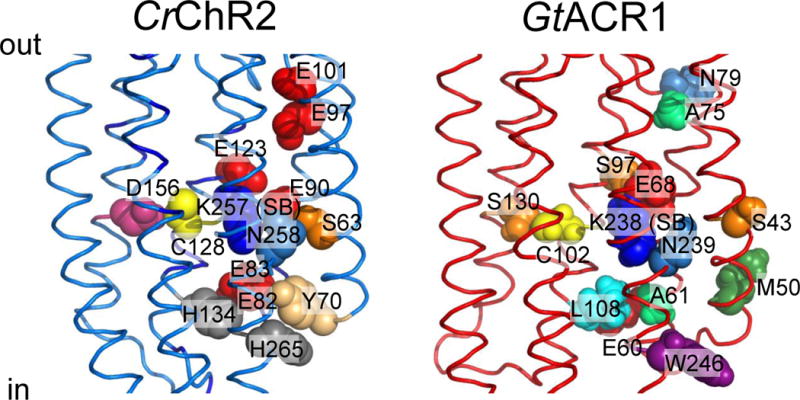
Microbial rhodopsins are a family of photoactive retinylidene proteins widespread throughout the microbial world. They are notable for their diversity of function, using variations of a shared seven-transmembrane helix design and similar photochemical reactions to carry out distinctly different light-driven energy and sensory transduction processes. Their study has contributed to our understanding of how evolution modifies protein scaffolds to create new protein chemistry, and their use as tools to control membrane potential with light is fundamental to optogenetics for research and clinical applications. We review the currently known functions and present more in-depth assessment of three functionally and structurally distinct types discovered over the past two years: (a) anion channelrhodopsins (ACRs) from cryptophyte algae, which enable efficient optogenetic neural suppression; (b) cryptophyte cation channelrhodopsins (CCRs), structurally distinct from the green algae CCRs used extensively for neural activation and from cryptophyte ACRs; and

Keywords: anion channelrhodopsins; cation channelrhodopsins; ion pumps; optogenetics; photosensors; retinylidene proteins.
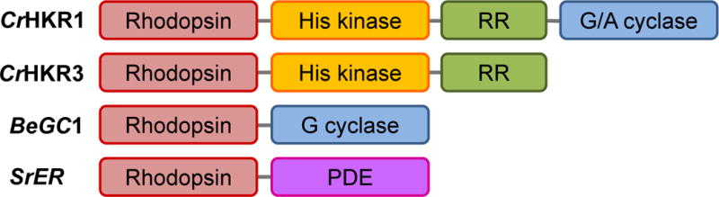

Figures

Figure 1
Figure 1
A cladogram of the microbial rhodopsin superfamily. For a list of sequences, accession numbers and source organisms see Supplementary Table 1.
Figure 2
Figure 2
Functional types of microbial rhodopsins. For molecules shown as ribbons high-resolution crystal structures have been obtained. Abbreviations: BRs, bacteriorhodopsins; PRs, proteorhodopsins; HRs, halorhodopsins; NaRs, Na+-pumping rhodopsins; SRs, sensory rhodopsins; ASR, Anabaena sensory rhodopsin; ER, enzymerhodopsins; CCRs, cation channelrhodopsins; ACRs, anion channelrhodopsins; eukar., eukaryotic; eubact., eubacterial; HK, histidine kinase; GC, gunanylyl cyclase; CD, cytoplasmic domain.
Figure 3
Figure 3
The domain structure of enzymerhodopsins. CrHKR1 and CrHKR3, histidine kinase 1 and 3, respectively, from the green alga C. reinhardtii; BeGC1, rhodopsin guanylylcyclase from the water mold B. emersonii; SrER, enzymerhodopsin from the choanoflagellate S. rosetta; RR, response regulator domain; G/A cyclase, gunanylyl/adenylyl cyclase domain.
Figure 4
Figure 4
Functionally important residues in chlorophyte CCRs and cryptophyte ACRs discussed in the text. Left, C1C2 crystal structure (3ug9) with residues numbered according to CrChR2 sequence. Right, GtACR1 homology model built using 3ug9 as a template. The side chains are colored according to their identity.
Figure 5
Figure 5
Helix 2 sequence logos of chlorophyte CCRs and cryptophyte ACRs created by WebLogo 3 as in (153). The overall height of each letter stack is proportional to the sequence conservation at that position, and the height of each letter is proportional to the frequency of the corresponding amino acid at that position. Acidic residues are red, and basic residues, blue. The residue numbers correspond to CrChR2 sequence.
Figure 6
Figure 6
Structural comparison of chlorophyte CCRs and ACRs. Gray, residues of the retinal binding pocket of BR conserved in each of the two types of channelrhodopsins; yellow, residues conserved in both CCRs and ACRs; blue, residues conserved only in CCRs; red, residues conserved only in ACRs. The residue conservation pattern is shown using the C1C2 crystal structure (3ug9; left) and a GtACR1 homology model built on the 3ug9 template (right).
Figure 7
Figure 7
The active site residues of cryptophyte CCRs. A GtCCR2 homology model built on the 2ksy template (middle) in comparison with those of the proton pump BR (1c3w; left) and chlorophyte CCR C1C2 (3ug9; right). For clarity only helices 3 and 7 are shown.

References

    1. Spudich JL, Yang C-S, Jung K-H, Spudich EN. Retinylidene proteins: structures and functions from archaea to humans. Annu Rev Cell Dev Biol. 2000;16:365–92. - PubMed
    1. Devine EL, Oprian DD, Theobald DL. Relocating the active-site lysine in rhodopsin and implications for evolution of retinylidene proteins. Proc Natl Acad Sci USA. 2013;110:13351–5. - PMC - PubMed
    1. Boyden ES. Optogenetics and the future of neuroscience. Nat Neurosci. 2015;18:1200–01. - PubMed
    1. Deisseroth K. Optogenetics: 10 years of microbial opsins in neuroscience. Nat Neurosci. 2015;18:1213–25. - PMC - PubMed
    1. Ernst OP, Lodowski DT, Elstner M, Hegemann P, Brown LS, Kandori H. Microbial and animal rhodopsins: structures, functions, and molecular mechanisms. Chem Rev. 2014;114:126–63. - PMC - PubMed

Publication types

MeSH terms