Review
. 2018 Feb 20;28(6):445-462.
doi: 10.1089/ars.2017.7057. Epub 2017 Apr 24.

The Role of Bacillithiol in Gram-Positive Firmicutes

Affiliations
Review

The Role of Bacillithiol in Gram-Positive Firmicutes

Pete Chandrangsu et al. Antioxid Redox Signal. .

Abstract

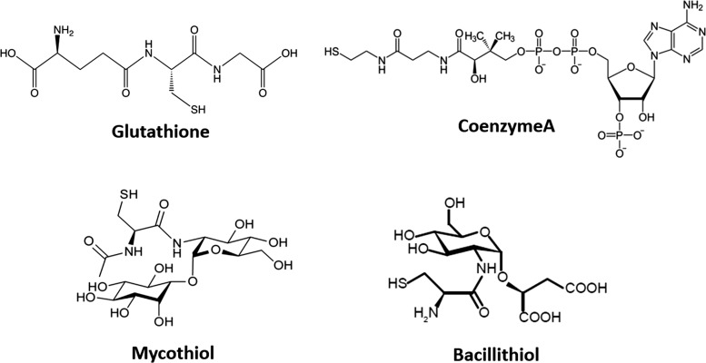
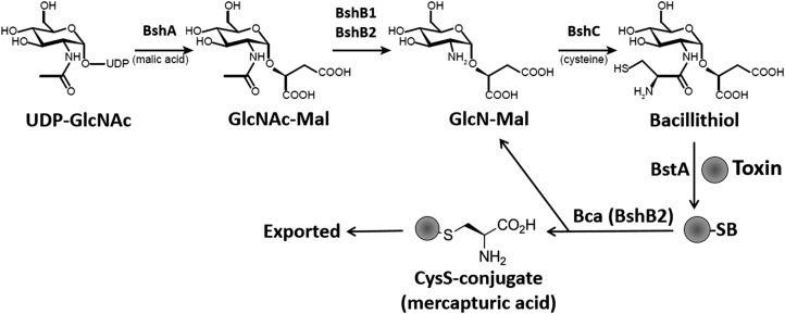
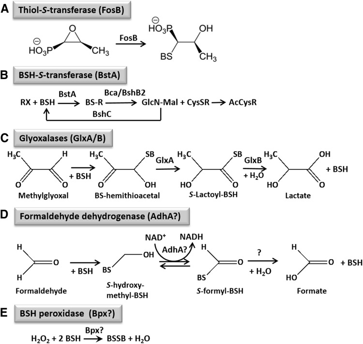
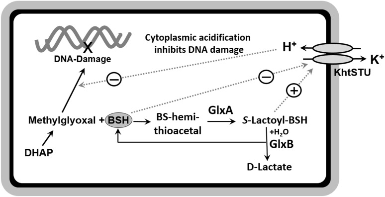
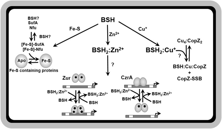
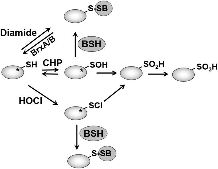
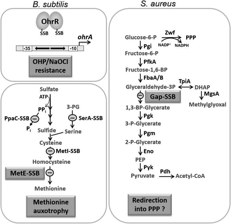
Significance: Since the discovery and structural characterization of bacillithiol (BSH), the biochemical functions of BSH-biosynthesis enzymes (BshA/B/C) and BSH-dependent detoxification enzymes (FosB, Bst, GlxA/B) have been explored in Bacillus and Staphylococcus species. It was shown that BSH plays an important role in detoxification of reactive oxygen and electrophilic species, alkylating agents, toxins, and antibiotics. Recent Advances: More recently, new functions of BSH were discovered in metal homeostasis (Zn buffering, Fe-sulfur cluster, and copper homeostasis) and virulence control in Staphylococcus aureus. Unexpectedly, strains of the S. aureus NCTC8325 lineage were identified as natural BSH-deficient mutants. Modern mass spectrometry-based approaches have revealed the global reach of protein S-bacillithiolation in Firmicutes as an important regulatory redox modification under hypochlorite stress. S-bacillithiolation of OhrR, MetE, and glyceraldehyde-3-phosphate dehydrogenase (Gap) functions, analogous to S-glutathionylation, as both a redox-regulatory device and in thiol protection under oxidative stress.

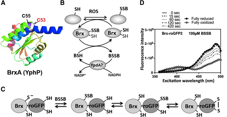
Critical issues: Although the functions of the bacilliredoxin (Brx) pathways in the reversal of S-bacillithiolations have been recently addressed, significantly more work is needed to establish the complete Brx reduction pathway, including the major enzyme(s), for reduction of oxidized BSH (BSSB) and the targets of Brx action in vivo.

Future directions: Despite the large number of identified S-bacillithiolated proteins, the physiological relevance of this redox modification was shown for only selected targets and should be a subject of future studies. In addition, many more BSH-dependent detoxification enzymes are evident from previous studies, although their roles and biochemical mechanisms require further study. This review of BSH research also pin-points these missing gaps for future research. Antioxid. Redox Signal. 28, 445-462.

Keywords: BSH biosynthesis; Bacillus subtilis; S-bacillithiolation; Staphylococcus aureus; bacilliredoxin; bacillithiol; metal homeostasis; methylglyoxal.


Figures

<b>FIG. 1.</b>
FIG. 1.
Structures of major LMW thiols in bacteria. Glutathione is utilized as the major LMW thiol in eukaryotes and Gram-negative bacteria, mycothiol in Actinomycetes, and BSH in Firmicutes. Coenzyme A (CoASH) also serves as an LMW thiol in Staphylococcus aureus and Bacillus anthracis. BSH, bacillithiol; LMW, low molecular weight.
<b>FIG. 2.</b>
FIG. 2.
Biosynthesis pathway of BSH and BSH-dependent detoxification. In the BSH synthesis pathway of Bacillus subtilis, the glycosyltransferase BshA first adds GlcNAc to malate-producing GlcNAc-Mal. Then, the paralog N-deacetylases BshB1 and BshB2 catalyze deacetylation of GlcNAc-Mal, and BshC adds cysteine (presumably in an unidentified activated form) to GlcN-Mal, producing BSH. Detoxification of toxins, xenobiotics, or electrophiles involves their conjugation with BSH by the BSH-S-transferase BstA, generating BS-conjugates that are cleaved by the deacetylase BshB2 (Bca) to CysS-conjugates and GlcN-Mal used for BSH recycling. The CysS-conjugates are exported from the cells as mercapturic acid derivatives. In S. aureus, only one BshB-like enzyme is present that functions both as deacetylase and amidase and is essential for BSH biosynthesis.
<b>FIG. 3.</b>
FIG. 3.
The functions of BSH-dependent detoxification enzymes in B. subtilis and S. aureus. BSH functions in detoxification of reactive oxygen and electrophilic species, HOCl, and antibiotics, such as fosfomycin in B. subtilis and S. aureus. (A) BSH is a cofactor for the thiol S-transferase FosB that adds BSH to fosfomycin for its detoxification. (B) Electrophiles, xenobiotics, and toxins (RX) are conjugated to BSH by the BSH S-transferase BstA to form BS-conjugates (BSR), which are cleaved by the BSH S-conjugate amidase BshB2 (Bca) to CysSR and a mercapturic acid (AcCySR) that is exported from the cell. (C) BSH functions in methylglyoxal detoxification as a cofactor for the glyoxalases I/II (GlxA/B) in B. subtilis. GlxA converts BS-hemithioacetal to S-lactoyl-BSH that is hydrolyzed by GlxB to D-lactate. (D) AdhA is a thiol-dependent formaldehyde dehydrogenase that is induced under FA stress (98), likely converting S-hydroxymethyl-BSH to S-formyl-BSH. In the final step, BSH and formate are released by an unidentified S-formyl-BSH hydrolase. (E) Unknown thiol-dependent peroxidases or peroxiredoxins (Bpx) might function in peroxide detoxification. Question marks indicate uncharacterized reactions.
<b>FIG. 4.</b>
FIG. 4.
BSH-dependent detoxification of methylglyoxal, leading to cytoplasmic acidification. Methylglyoxal can be produced as a byproduct of the glycolysis from DHAP. Methylglyoxal reacts spontaneously with BSH, forming BS-hemithioacetal, which is converted to S-lactoyl-BSH by the glyoxalase-I (GlxA) and to lacatate by the glyoxalase-II (GlxB). S-lactoyl-BSH activates the potassium proton antiporter KhtSTU for K-efflux and proton import, leading to cytoplasmic acidification that likely inhibits interaction of methylglyoxal with the DNA to prevent DNA damage. BSH inhibits the antiporter KhtSTU. DHAP, dihydroxyacetone phosphate.
<b>FIG. 5.</b>
FIG. 5.
The functions of BSH in metal homeostasis. A role for BSH in metal homeostasis has been identified for iron, zinc, and copper. BSH is required for the full activity of FeS requiring proteins in S. aureus, possibly as a key component in the assembly and delivery of FeS clusters. BSH is believed to function in an independent, yet overlapping role with the FeS carrier proteins, SufA and Nfu. BSH can also bind zinc with a high affinity and serves as a major cytosolic zinc buffer as demonstrated in B. subtilis, allowing the cell to avoid zinc intoxication under conditions of excess. Biochemical evidence also suggests a role for BSH in facilitating the delivery and removal of zinc from the zinc-sensing metalloregulators, Zur and CzrA. Lastly, BSH may also work in concert with CopZ in interacellular copper buffering and delivery to metalloproteins and may protect CopZ from overoxidation through S-bacilliothiolation. FeS, iron-sulfur.
<b>FIG. 6.</b>
FIG. 6.
Mechanisms of S-bacillithiolation and its reversal. Proteins are S-bacillithiolated under CHP, HOCl, and diamide stress in Bacillus and Staphylococcus species. Diamide is a reactive electrophile species leading directly to the formation of mixed BSH disulfides. CHP and HOCl activate thiols to a sulfenic acid (-SOH) and sulfenylchloride (-SCl) intermediates, respectively, that react further with BSH to form S-bacillithiolated proteins. In the absence of proximal thiols, -SOH and -SCl are overoxidized to sulfinic or sulfonic acids. Thus, S-bacillithiolation serves to protect vulnerable thiols against irreversible overoxidation. The asterisk indicates that often active site Cys residues are targets for S-bacillithiolation. The reversal of S-bacillithiolation is catalyzed by the BrxA/B. Brx, bacilliredoxin; CHP, cumene hydroperoxide.
<b>FIG. 7.</b>
FIG. 7.
Physiological roles of S-bacillithiolations for OhrR and MetE in B. subtilis and for Gap of S. aureus. NaOCl leads to S-bacillithiolation of OhrR and MetE as main targets in B. subtilis that have regulatory roles under NaOCl stress. S-bacillithiolation inactivates the OhrR repressor, leading to induction of the OhrA peroxiredoxin that confers resistance to NaOCl and OHP. S-bacillithiolation of the methionine synthase MetE at its active site Cys730 and of other enzymes of the Cys and Met biosynthesis pathway (YxjG, PpaC, SerA, MetI) leads to methionine auxotrophy. In S. aureus, the glycolytic Gap is the main target for S-bacillithiolation under NaOCl stress. S-bacillithiolation of the Gap active site Cys151 leads to reversible Gap inactivation and prevents its overoxidation to Cys sulfonic acid. Gap inactivation under oxidative stress might redirect the glycolytic flux into the PPP for NADPH regeneration, as shown in yeast cells. Gap, glyceraldehyde-3-phosphate dehydrogenase; OHP, organic hydroperoxide; PPP, pentose phosphate pathway.
<b>FIG. 8.</b>
FIG. 8.
Structure of the BrxA (YphP) of B. subtilis (A), Brx redox pathway (B), principle of Brx-roGFP2 biosensor oxidation (C), and ratiometric change of excitation spectra of Brx-roGFP2 by BSSB in vitro (D). (A) The structure of the BrxA (YphP) with the CGC active site motif was generated by using the software Phyre2 and PyMol. (B) BrxA reduces S-bacillithiolated proteins, resulting in Brx-SSB formation. Recycling of BrxA may require BSH and an NADPH-dependent BSSB reductase that could be YpdA. (C) The Brx-roGFP2 biosensor reacts first with BSSB at the active site Cys of Brx, leading to Brx-SSB formation, subsequent transfer of the BSH moiety to the coupled roGFP2, and re-arrangement to the roGFP2 disulfide. The roGFP2 disulfide causes a change of the 405/488 nm excitation ratio. (D) Brx-roGFP2 reacts very fast with purified 100 μM BSSB, as shown by the ratiometric change in the excitation maxima at 405 and 488 nm. For fully oxidized and reduced probes, Brx-roGFP2 was treated with 5 mM diamide and 10 mM DTT, respectively. The Brx-roGFP2 fluorescence excitation spectra were monitored by using the Clariostar microplate reader. Adapted from a previous publication (73). BSSB, oxidized bacillithiol disulfide.

References

    1. Akanuma G, Nanamiya H, Natori Y, Nomura N, and Kawamura F. Liberation of zinc-containing L31 (RpmE) from ribosomes by its paralogous gene product, YtiA, in Bacillus subtilis. J Bacteriol 188: 2715–2720, 2006 - PMC - PubMed
    1. Allen EM. and Mieyal JJ. Protein-thiol oxidation and cell death: regulatory role of glutaredoxins. Antioxid Redox Signal 17: 1748–1763, 2012 - PMC - PubMed
    1. Andreini C, Banci L, Bertini I, and Rosato A. Zinc through the three domains of life. J Proteome Res 5: 3173–3178, 2006 - PubMed
    1. Baichoo N, Wang T, Ye R, and Helmann JD. Global analysis of the Bacillus subtilis Fur regulon and the iron starvation stimulon. Mol Microbiol 45: 1613–1629, 2002 - PubMed
    1. Banci L, Bertini I, Del Conte R, Markey J, and Ruiz-Duenas FJ. Copper trafficking: the solution structure of Bacillus subtilis CopZ. Biochemistry 40: 15660–15668, 2001 - PubMed

Publication types

MeSH terms

LinkOut - more resources