

Population-based study of the incidence of congenital hip dysplasia in preterm infants from the Survey of Neonates in Pomerania (SNiP)
- PMID: 28302080
- PMCID: PMC5356283
- DOI: 10.1186/s12887-017-0829-5
Population-based study of the incidence of congenital hip dysplasia in preterm infants from the Survey of Neonates in Pomerania (SNiP)
Abstract
Background: Some etiological factors involved in developmental dysplasia of the hip (DDH) occur in the last trimester of pregnancy, which could result in a decreased incidence of DDH in preterm infants. The aim of this study was to compare the incidence of DDH between preterm and term infants.
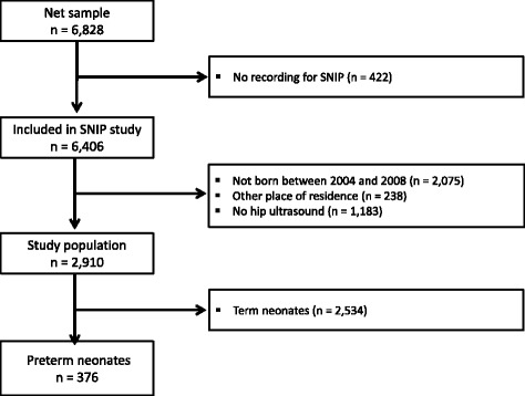
Methods: Ultrasound of the hip joint was performed in 2,534 term infants and 376 preterm infants within the population-based Survey of Neonates in Pomerania (SNiP) study.
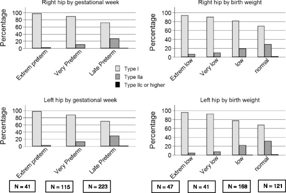
Results: A total of 42 (1.66%) term infants had DDH (Graf type II c, 0.8%; type D, 0.3% left and 0.4% right; type III a, 0.2% left). Eighteen infants had bilateral findings. Hip dysplasia occurred more frequently in female neonates (32/1,182 vs. 10/1,302, p < 0.023; 95% CI 0.012-0.022, χ 2 test). A familial disposition for DDH was found in 169 (6.7%) term infants and 181 (7.1%) infants in the overall population. In preterm infants, dysplasia of the hip was found in only three late preterm infants with gestational age between 36 and 37 weeks (n = 97) and not in preterm infants <36 weeks gestational age (n = 279). Regression analysis revealed a narrowly significant association between gestational week of birth and DDH (relative risk = 1.17; 95% confidence interval 0.99-1.37; p = 0.065).
Conclusion: Our study suggests that preterm infants <36 weeks gestational age have a decreased risk of DDH.
Keywords: Hip dysplasia; Preterm neonate; Screening of neonates; Ultrasound of the hip.
Figures

Similar articles
-
Breech preterm infants are at risk of developmental dysplasia of the hip.J Paediatr Child Health. 2013 Aug;49(8):658-63. doi: 10.1111/jpc.12250. Epub 2013 Jun 12. J Paediatr Child Health. 2013. PMID: 23758088
-
Developmental dysplasia of the hip in preterm breech infants.Arch Dis Child Fetal Neonatal Ed. 2020 Sep;105(5):556-558. doi: 10.1136/archdischild-2019-317658. Epub 2020 Jan 3. Arch Dis Child Fetal Neonatal Ed. 2020. PMID: 31900256
-
Cochrane Review: Screening programmes for developmental dysplasia of the hip in newborn infants.Evid Based Child Health. 2013 Jan;8(1):11-54. doi: 10.1002/ebch.1891. Evid Based Child Health. 2013. PMID: 23878122 Review.
-
Sonographic screening for developmental dysplasia of the hip in preterm breech infants: do current guidelines address the specific needs of premature infants?J Perinatol. 2016 Jul;36(7):552-6. doi: 10.1038/jp.2016.7. Epub 2016 Feb 25. J Perinatol. 2016. PMID: 26914014
-
Is prematurity a risk factor for developmental dysplasia of the hip? A systematic review and meta-analysis.J Pediatr Orthop B. 2023 Jul 1;32(4):305-311. doi: 10.1097/BPB.0000000000001021. Epub 2022 Nov 14. J Pediatr Orthop B. 2023. PMID: 36445370
Cited by
-
Trends in the Prevalences of Selected Birth Defects in Korea (2008⁻2014).Int J Environ Res Public Health. 2018 May 5;15(5):923. doi: 10.3390/ijerph15050923. Int J Environ Res Public Health. 2018. PMID: 29734759 Free PMC article.
-
Universal Clinical DDH Screening Complemented with Targeted Ultrasound Is Effective in Finland.J Bone Joint Surg Am. 2025 Apr 2;107(7):e26. doi: 10.2106/JBJS.24.00313. Epub 2025 Feb 20. J Bone Joint Surg Am. 2025. PMID: 39977488 Free PMC article.
-
Sonographic Hip Angles in Relation to Gestational Age of Neonates. A Prospective, Cohort in the Population of Northern Greece.Arch Bone Jt Surg. 2023;11(3):197-205. doi: 10.22038/ABJS.2022.67942.3219. Arch Bone Jt Surg. 2023. PMID: 37168591 Free PMC article.
-
The Global Research Trends and Hotspots on Developmental Dysplasia of the Hip: A Bibliometric and Visualized Study.Front Surg. 2021 Oct 25;8:671403. doi: 10.3389/fsurg.2021.671403. eCollection 2021. Front Surg. 2021. PMID: 34760913 Free PMC article.
-
Incidence of Neonatal Developmental Dysplasia of the Hip and Late Detection Rates Based on Screening Strategy: A Systematic Review and Meta-analysis.JAMA Netw Open. 2022 Aug 1;5(8):e2227638. doi: 10.1001/jamanetworkopen.2022.27638. JAMA Netw Open. 2022. PMID: 35980635 Free PMC article.
References
Publication types
MeSH terms
LinkOut - more resources
Full Text Sources
Other Literature Sources
Medical
Research Materials
