In vivo confocal microscopy of pre-Descemet corneal dystrophy associated with X-linked ichthyosis: a case report
- PMID: 28302098
- PMCID: PMC5356324
- DOI: 10.1186/s12886-017-0423-5
In vivo confocal microscopy of pre-Descemet corneal dystrophy associated with X-linked ichthyosis: a case report
Abstract
Background: Pre-Descemet corneal dystrophy (PDCD) is characterized by the presence of numerous, tiny, polymorphic opacities immediately anterior to Descemet membrane, which is a rare form of corneal stromal dystrophy and hard to be diagnosed. In vivo confocal microscopy (IVCM) is a useful tool to examine the minimal lesions of the cornea at the cellular level. In this article, we report a rare case of PDCD associated with X-linked ichthyosis and evaluate IVCM findings.
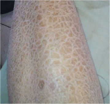
Case presentation: We present a 34-year-old male Chinese patient with PDCD associated with X-linked ichthyosis. Slit-lamp biomicroscopy showed the presence of tiny and pleomorphic opacities in the posterior stroma immediately anterior to Descemet membrane bilaterally. IVCM revealed regular distributed hyperreflective particles inside the enlarged and activated keratocytes in the posterior stroma. Hyperreflective particles were also observed dispersedly outside the keratocytes in the anterior stroma. Dermatological examination revealed that the skin over the patient's entire body was dry and coarse, with thickening and scaling of the skin in the extensor side of the extremities. PCR results demonstrated that all ten exons and part flanking sequences of STS gene failed to produce any amplicons in the patient.
Conclusions: IVCM is useful for analyzing the living corneal structural changes in rare corneal dystrophies. We first reported the IVCM characteristics of PDCD associated with X-linked ichthyosis, which was caused by a deletion of the steroid sulfatase (STS) gene, confirmed by gene analysis.
Keywords: In vivo confocal microscopy; Pre-Descemet corneal dystrophy; Steroid sulfatase; X-linked ichthyosis.
Figures


References
-
- Chen PL, Tang KP, Liang JB. Pre-Descemet's corneal dystrophy associated with ichthyosis. Chinese Medical Journal (Taipei) 2002;65:407–409. - PubMed
-
- Malhotra C, Jain AK, Dwivedi S, Chakma P, Rohilla V, Sachdeva K. Characteristics of Pre-Descemet Membrane Corneal Dystrophy by Three Different Imaging Modalities-In Vivo Confocal Microscopy, Anterior Segment Optical Coherence Tomography, and Scheimpflug Corneal Densitometry Analysis. Cornea. 2015;34:829–832. doi: 10.1097/ICO.0000000000000454. - DOI - PubMed
Publication types
MeSH terms
LinkOut - more resources
Full Text Sources
Other Literature Sources

