

Spinal epidural lipomatosis - an easily ignored secondary intraspinal disorder in spinal kyphotic deformities
- PMID: 28302104
- PMCID: PMC5356279
- DOI: 10.1186/s12891-017-1467-7
Spinal epidural lipomatosis - an easily ignored secondary intraspinal disorder in spinal kyphotic deformities
Abstract
Background: A previous study reported a high prevalence of spinal epidural lipomatosis (SEL) in patients with Scheuermann kyphosis (SK) and suggested that it may play a role in the pathogenesis of this disease. According to our observation, however, SEL occurs in other spinal kyphotic deformities as well. The aim of this study was to test the hypothesis that SEL commonly occurs in patients with different types of kyphotic deformities as a secondary intraspinal disorder.
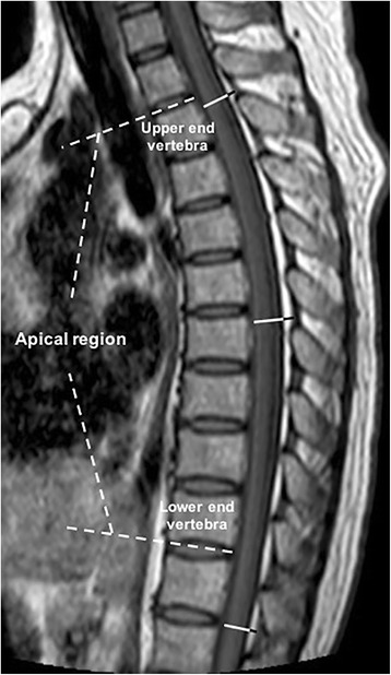
Methods: MR images of 16 patients with congenital kyphosis (CK), 40 patients with SK, 13 patients with tuberculotic kyphosis (TK), and 69 age- and sex-matched controls were retrospectively evaluated. The body mass index (BMI), kyphosis Cobb angle, and sagittal diameters of spinal epidural fat (EF) and the dural sac (DS) in the apical region (EFA, DSA) and non-kyphotic region (EFN, DSN) were measured. The EF ratios at the apical vertebral level (EFRA) and in the non-kyphotic region (EFRN) were calculated as EF / (EF + DS).
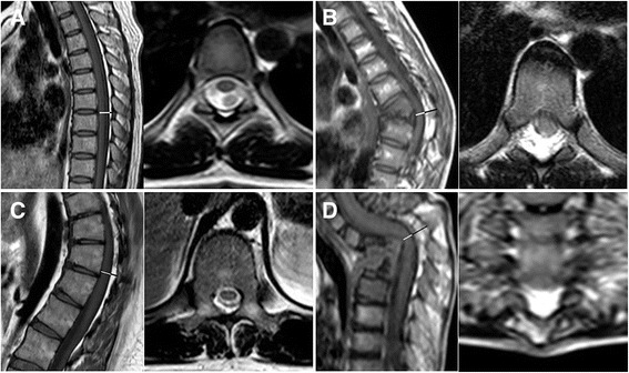
Results: EFA and EFRA were significantly higher among patients with CK, SK, and TK than among controls (P < 0.05). Seven CK patients (43.8%), 8 SK patients (20.0%), and 11 TK patients (84.6%) fulfilled the diagnostic criteria for SEL, while only 6.3, 2.5, and 0% of patients in the control groups did (P = 0.019, 0.014, and < 0.001, respectively). Spearman's correlation analysis showed statistically significant correlations between the kyphosis Cobb angle and the amount of EF in all three patient groups.
Conclusions: SEL is a common secondary intraspinal disorder in different types of kyphotic deformities, and surgeons should pay increased attention to this intraspinal anomaly because excessive EF may compress the spinal cord and cause neurological deficits.
Keywords: Congenital kyphosis; Scheuermann kyphosis; Spinal epidural lipomatosis; Spinal kyphotic deformities; Tuberculotic kyphosis.
Figures
Similar articles
-
Surgical management of thoracic myelopathy from long-segment epidural lipomatosis with skip hemilaminotomies: illustrative case.J Neurosurg Case Lessons. 2021 Dec 13;2(24):CASE21595. doi: 10.3171/CASE21595. eCollection 2021 Dec 13. J Neurosurg Case Lessons. 2021. PMID: 35855484 Free PMC article.
-
Spinal epidural lipomatosis: a common imaging feature in Scheuermann disease.J Spinal Disord Tech. 2012 Oct;25(7):356-61. doi: 10.1097/BSD.0b013e31822631d3. J Spinal Disord Tech. 2012. PMID: 21705916
-
MRI Screening in Operative Scheuermann Kyphosis: Is it Necessary?Spine Deform. 2017 Mar;5(2):124-133. doi: 10.1016/j.jspd.2016.10.008. Spine Deform. 2017. PMID: 28259264
-
Decompression of idiopathic lumbar epidural lipomatosis: diagnostic magnetic resonance imaging evaluation and review of the literature.J Neurosurg Spine. 2006 Jan;4(1):24-30. doi: 10.3171/spi.2006.4.1.24. J Neurosurg Spine. 2006. PMID: 16506462 Review.
-
Management of idiopathic spinal epidural lipomatosis: a case report and review of the literature.Childs Nerv Syst. 2018 Apr;34(4):757-763. doi: 10.1007/s00381-017-3706-5. Epub 2017 Dec 22. Childs Nerv Syst. 2018. PMID: 29273822 Review.
Cited by
-
Surgical management of thoracic myelopathy from long-segment epidural lipomatosis with skip hemilaminotomies: illustrative case.J Neurosurg Case Lessons. 2021 Dec 13;2(24):CASE21595. doi: 10.3171/CASE21595. eCollection 2021 Dec 13. J Neurosurg Case Lessons. 2021. PMID: 35855484 Free PMC article.
-
Could Spinal Epidural Lipomatosis Be the Hallmark of Metabolic Syndrome on the Spine? A Literature Review with Emphasis on Etiology.Diagnostics (Basel). 2023 Jan 16;13(2):322. doi: 10.3390/diagnostics13020322. Diagnostics (Basel). 2023. PMID: 36673132 Free PMC article. Review.
-
Spinal epidural lipomatosis in a pediatric patient with a malignant brain tumor: illustrative case.J Neurosurg Case Lessons. 2023 Feb 20;5(8):CASE22556. doi: 10.3171/CASE22556. Print 2023 Feb 20. J Neurosurg Case Lessons. 2023. PMID: 36806011 Free PMC article.
References
-
- Nakahara D, Yonezawa I, Kobanawa K, Sakoda J, Nojiri H, Kamano S, Okuda T, Kurosawa H. Magnetic resonance imaging evaluation of patients with idiopathic scoliosis: a prospective study of four hundred seventy-two outpatients. Spine. 2011;36(7):E482–485. doi: 10.1097/BRS.0b013e3181e029ed. - DOI - PubMed
MeSH terms
LinkOut - more resources
Full Text Sources
Other Literature Sources
Research Materials
