

[Effects of ω-3 polyunsaturated fatty acids on lymphocyte apoptosis rate in rats with sepsis]
- PMID: 28302212
- PMCID: PMC7390154
- DOI: 10.7499/j.issn.1008-8830.2017.03.021
[Effects of ω-3 polyunsaturated fatty acids on lymphocyte apoptosis rate in rats with sepsis]
Abstract
Objective: To investigate the effects of ω-3 polyunsaturated fatty acids (ω-3 PUFAs) on the apoptosis of thymic and splenic lymphocytes in rats with sepsis.
Methods: A total of 80 female Sprague-Dawley rats aged 7-8 weeks were randomly divided into model group, conventional lipid emulsion group (0.1 g/kg daily), low-dose ω-3 PUFAs group (0.1 g/kg daily), middle-dose ω-3 PUFAs group (0.2 g/kg daily), and high-dose ω-3 PUFAs group (0.3 g/kg daily). Cecal ligation and puncture were used to establish a rat model of sepsis. The treatment groups were then given tail vein injection of lipid emulsion or glucose diluents of ω-3 PUFAs at different doses, and the model group was given glucose injection via the tail vein at the same dose. According to the time of sacrifice, each group was further divided into 24-hour and 72-hour subgroups, with 8 rats in each subgroup. Hematoxylin and eosin staining was used to observe the pathological changes in the thymus and spleen. TUNEL was used to measure the apoptosis rates of thymic and splenic lymphocytes.
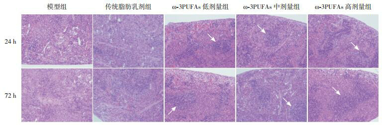
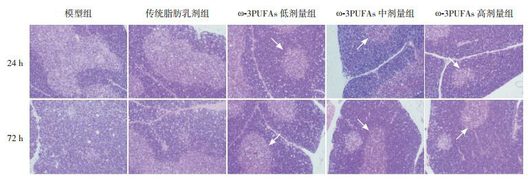
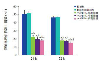
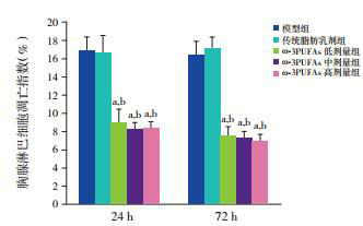
Results: In the three ω-3 PUFAs groups, the rats had a complete thymic lobular structure and clear structures of the cortex and medulla. In the model and the conventional lipid emulsion groups, the boundaries of the cortex and medulla were unclear and the number of lymphocytes was significantly reduced. In the ω-3 PUFAs groups, the structure of the red and white pulp of the spleen was maintained with the presence of splenic follicles, while in the model and the conventional lipid emulsion groups, the structure of the red and white pulp of the spleen was disordered and splenic follicles were significantly reduced or disappeared. Compared with the model and the conventional lipid emulsion groups, the ω-3 PUFAs groups showed significant reductions in the apoptosis rates of thymic and splenic lymphocytes at 24 and 72 hours (P<0.01). Compared with the low-dose ω-3 PUFAs group, the high-dose ω-3 PUFAs group had significantly reduced apoptosis rates of splenic lymphocytes at 24 and 72 hours (P<0.05).
Conclusions: ω-3 PUFAs can reduce the apoptosis of thymic and splenic lymphocytes in rats with sepsis in a dose-dependent manner.
目的: 探讨ω-3多不饱和脂肪酸对脓毒症大鼠胸腺和脾脏淋巴细胞凋亡的影响。
方法: 将80只7~8周龄雌性Sprague-Dawley大鼠随机分成模型组、传统脂肪乳剂治疗组(每日0.1 g/kg)、ω-3PUFAs低剂量治疗组(每日0.1 g/kg)、ω-3PUFAs中剂量治疗组(每日0.2 g/kg)和ω-3PUFAs高剂量治疗组(每日0.3 g/kg)。采用盲肠结扎穿孔法建立脓毒症大鼠模型,造模后各治疗组分别给予脂肪乳剂和不同剂量ω-3PUFAs葡萄糖稀释液尾静脉注射给药,模型组予等量葡萄糖注射液尾静脉注射。按处死时间将各组大鼠随机分为24 h和72 h两个亚组,每组8只大鼠。采用苏木精-伊红染色观察各组大鼠胸腺和脾脏的病理改变;采用TUNEL法检测各组大鼠脾脏和胸腺淋巴细胞凋亡指数。
结果: 不同剂量ω-3PUFAs组大鼠胸腺小叶结构完整,皮髓质结构清晰,模型组和传统脂肪乳剂治疗组皮髓质界限不清,淋巴细胞明显减少;ω-3PUFAs组脾脏红髓和白髓结构尚能保持,可见脾小结存在,而模型组和传统脂肪乳剂治疗组红髓和白髓结构紊乱,脾小结明显缩小或消失。不同剂量ω-3PUFAs组24 h和72 h胸腺和脾脏淋巴细胞凋亡率均较模型组、传统脂肪乳剂治疗组显著降低(P < 0.01),ω-3PUFAs高剂量治疗组24 h和72 h脾脏淋巴细胞凋亡率均较ω-3PUFAs低剂量治疗组显著降低(P < 0.05)。
结论: ω-3多不饱和脂肪酸有减轻脓毒症大鼠胸腺和脾脏淋巴细胞凋亡的作用,并且可能与剂量相关。
Figures
References
-
- Moore LJ, McKinley BA, Turner KL, et al. The epidemiology of sepsis in general surgery patients. J Trauma. 2011;70(3):672–680. - PubMed
-
- de Souza DC, Barreira ER, Faria LS. The epidemiology of sepsis in childhood. Shock. 2017;47(1S Suppl 1):2–5. - PubMed
-
- de Souza DC, Shieh HH, Barreira ER, et al. Epidemiology of sepsis in children admitted to PICUs in South America. https://www.researchgate.net/publication/304668737_Epidemiology_of_Sepsi.... Pediatr Crit Care Med. 2016;17(8):727–734. - PubMed
MeSH terms
Substances
LinkOut - more resources
Full Text Sources
Medical