

Genetically introduced hydrogen bond interactions reveal an asymmetric charge distribution on the radical cation of the special-pair chlorophyll P680
- PMID: 28302724
- PMCID: PMC5418047
- DOI: 10.1074/jbc.M117.781062
Genetically introduced hydrogen bond interactions reveal an asymmetric charge distribution on the radical cation of the special-pair chlorophyll P680
Abstract
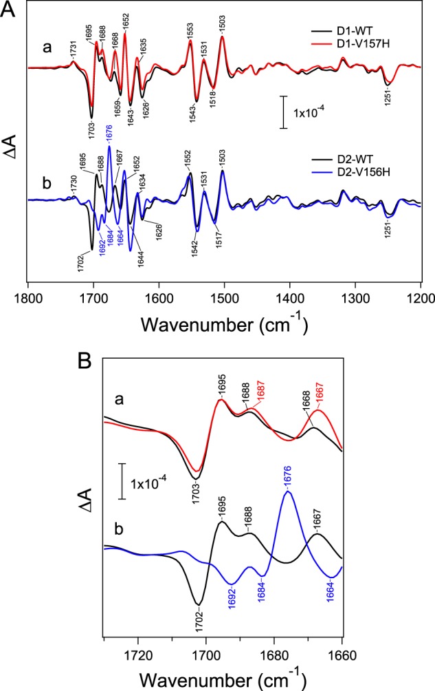
The special-pair chlorophyll (Chl) P680 in photosystem II has an extremely high redox potential (Em ) to enable water oxidation in photosynthesis. Significant positive-charge localization on one of the Chl constituents, PD1 or PD2, in P680+ has been proposed to contribute to this high Em To identify the Chl molecule on which the charge is mainly localized, we genetically introduced a hydrogen bond to the 131-keto C=O group of PD1 and PD2 by changing the nearby D1-Val-157 and D2-Val-156 residues to His, respectively. Successful hydrogen bond formation at PD1 and PD2 in the obtained D1-V157H and D2-V156H mutants, respectively, was monitored by detecting 131-keto C=O vibrations in Fourier transfer infrared (FTIR) difference spectra upon oxidation of P680 and the symmetrically located redox-active tyrosines YZ and YD, and they were simulated by quantum-chemical calculations. Analysis of the P680+/P680 FTIR difference spectra of D1-V157H and D2-V156H showed that upon P680+ formation, the 131-keto C=O frequency upshifts by a much larger extent in PD1 (23 cm-1) than in PD2 (<9 cm-1). In addition, thermoluminescence measurements revealed that the D1-V157H mutation increased the Em of P680 to a larger extent than did the D2-V156H mutation. These results, together with the previous results for the mutants of the His ligands of PD1 and PD2, lead to a definite conclusion that a charge is mainly localized to PD1 in P680<sup/>.
Keywords: FTIR; chlorophyll; cyanobacteria; hydrogen bond; photosynthesis; photosystem II; quantum chemical calculation; site-directed mutagenesis; special pair; thermoluminescence.
© 2017 by The American Society for Biochemistry and Molecular Biology, Inc.
Conflict of interest statement
The authors declare that they have no conflicts of interest with the contents of this article
Figures


References
-
- Debus R. J. (1992) The manganese and calcium ions of photosynthetic oxygen evolution. Biochim. Biophys. Acta 1102, 269–352 - PubMed
-
- McEvoy J. P., and Brudvig G. W. (2006) Water-splitting chemistry of photosystem II. Chem. Rev. 106, 4455–4483 - PubMed
-
- Messinger J., Noguchi T., and Yano J. (2012) in Molecular Solar Fuels (Wydrzynski T. J., and Hillier W., eds) pp. 163–207, Royal Society of Chemistry, Cambridge, UK
-
- Grundmeier A., and Dau H. (2012) Structural models of the manganese complex of photosystem II and mechanistic implications. Biochim. Biophys. Acta 1817, 88–105 - PubMed
-
- Shen J.-R. (2015) The structure of photosystem II and the mechanism of water oxidation in photosynthesis. Annu. Rev. Plant Biol. 66, 23–48 - PubMed
Publication types
MeSH terms
Substances
Associated data
- Actions
LinkOut - more resources
Full Text Sources
Other Literature Sources
