Review
. 2017 Mar 17;120(6):941-959.
doi: 10.1161/CIRCRESAHA.116.309040.

Cardiac Regeneration: Lessons From Development

Affiliations
Review

Cardiac Regeneration: Lessons From Development

Francisco X Galdos et al. Circ Res. .

Abstract

Palliative surgery for congenital heart disease has allowed patients with previously lethal heart malformations to survive and, in most cases, to thrive. However, these procedures often place pressure and volume loads on the heart, and over time, these chronic loads can cause heart failure. Current therapeutic options for initial surgery and chronic heart failure that results from failed palliation are limited, in part, by the mammalian heart's low inherent capacity to form new cardiomyocytes. Surmounting the heart regeneration barrier would transform the treatment of congenital, as well as acquired, heart disease and likewise would enable development of personalized, in vitro cardiac disease models. Although these remain distant goals, studies of heart development are illuminating the path forward and suggest unique opportunities for heart regeneration, particularly in fetal and neonatal periods. Here, we review major lessons from heart development that inform current and future studies directed at enhancing cardiac regeneration.

Keywords: cardiac development; cardiac regeneration; cardiomyocyte maturation; signaling pathways; transcriptional regulation.


Conflict of interest statement

Conflict of Interest

None

Figures

Figure 1
Figure 1. Specification of mesodermal precursors
Schematic representing the signaling events leading to mesodermal specification during early development. NODAL is first expressed proximally at E5.0. Through an autoregulatory loop, NODAL activates its own expression throughout the epiblast (shown in light purple) and goes on to induce the expression of NODAL antagonists, LEFTY1 and CER1 in the distal visceral endoderm at E5.5 (DVE). The DVE migrates anteriorly where it specifies the anterior portion of the embryo as shown in the yellow hues at E6.5-7.5. The anterior visceral endoderm (AVE, yellow) limits NODAL signaling to the posterior of the embryo. Along with WNT3 and BMP signaling, NODAL specifies early primitive streak progenitors to the mesoderm fate.
Figure 2
Figure 2. Regulation of cardiac progenitor proliferation and differentiation
A. Schematic showing the anterolateral position of first heart field (FHF) progenitors and dorsomedial position of second heart field (SHF) progenitors at E7.5. Canonical WNTs, Sonic Hedgehog (SHH), and fibroblast growth factors (FGFs) are expressed dorsally in the region encompassed by the SHF, while non-canonical WNTs, BMP2, and FGF8 are expressed ventrally, where the FHF is present. FHF progenitors make up the cardiac crescent and differentiate prior to the SHF in order to form the developing heart tube at E8.0. SHF maintain their proliferative state and elongate the heart tube by migrating and differentiating at the inflow and outflow poles of the heart. B. Non-canonical WNTs, BMP2/4, and FGF8 signaling drives FHF progenitors to differentiates towards the myocyte lineage. Meanwhile, Canonical WNT/β-catenin, SHH, and FGFs maintain SHF progenitor proliferation. SHF progenitor migration to the outflow and inflow poles of the heart tube exposes them to BMP2/4 and non-canonical WNTs which drives SHF progenitors to exit their proliferative state and differentiate. C) Canonical WNT/ β-catenin signaling inhibits the differentiation of cardiac progenitors to the myocytes. BMP signaling activates SMAD4 which binds to the transcription factor HOPX to directly inhibit Canonical WNT/β-catenin. Moreover, non-canonical WNTs such as WNT5a and WNT11 also inhibit Canonical WNT/β-catenin to drive cardiac progenitor differentiation. (Illustration credit: Ben Smith)
Figure 3
Figure 3. Model of the trabeculation process
During trabeulation, a small fraction of CMs in the compact myocardium (pink) are first specified as trabeculating CMs (brown). These cells delaminate from the compact myocardium and migrate inward to form the first trabecular CMs. CMs in both compacted and trabecular myocardium further proliferate. This proliferation, together with CM migration and rearrangement, results in protrusion and expansion of the trabecular myocardium. (Illustration Credit: Ben Smith)
Figure 4
Figure 4. Model of cardiomyocyte regeneration
During heart regeneration, myocardial injury and CM loss induce the dedifferentiation of a fraction of CMs (pink). These CMs reenter the cell cycle, produce new CMs, and replenish the lost CMs. CMs that are generated from cell division re-differentiate to a fully mature state to improve heart contraction.
Figure 5
Figure 5. Representative cellular events during CM maturation
During CM maturation, transcriptional changes occur throughout both embryonic and postnatal development. CMs stop hyperplastic growth at the perinatal stage and transition to hypertrophic growth, in which sarcomere number increases and sarcomere alignment improves. Altered expression of critical sarcomere isoforms modifies the contractile properties of CMs. Mitochondria increase in number and size and become well organized with respect to sarcomeres. After birth, CMs switch from deriving most of their energy from glycolysis to depending upon oxidative phosphorylation. The maturation of electrophysiologic properties is characterized by proper expression and localization of ion channels characteristic of adult CMs. T-tubules form at a late stage of postnatal development, perhaps to permit rapid AP penetration with rapidly enlarging CMs. Maturation is also characterized by the postnatal establishment of intercalated discs and proper formation of cell-cell contacts.

References

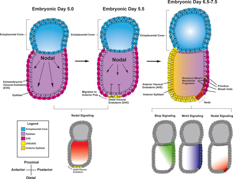
    1. Arnold SJ, Robertson EJ. Making a commitment: cell lineage allocation and axis patterning in the early mouse embryo. Nature Reviews Molecular Cell Biology. 2009;10:91–103. - PubMed
    1. Brennan J, Lu CC, Norris DP, Rodriguez TA. Nodal signalling in the epiblast patterns the early mouse embryo. Nodal signalling in the epiblast patterns the early mouse embryo. 2001 - PubMed
    1. Waldrip WR, Bikoff EK, Hoodless PA, Wrana JL, Robertson EJ. Smad2 signaling in extraembryonic tissues determines anterior-posterior polarity of the early mouse embryo. Cell. 1998;92:797–808. - PubMed
    1. Perea-Gomez A, Vella F, Shawlot W, Oulad-Abdelghani M, Chazaud C, Meno C, Pfister V, Chen L, Robertson E, Hamada H, Behringer RR, Ang S-L. Nodal Antagonists in the Anterior Visceral Endoderm Prevent the Formation of Multiple Primitive Streaks. Developmental Cell. 2002;3:745–756. - PubMed
    1. Conlon FL, Lyons KM, Takaesu N, Barth KS, Kispert A, Herrmann B, Robertson EJ. A primary requirement for nodal in the formation and maintenance of the primitive streak in the mouse. Development (Cambridge, England) 1994;120:1919–1928. - PubMed