. 2017 Mar 17;49(3):e302.
doi: 10.1038/emm.2016.167.

TMF and glycitin act synergistically on keratinocytes and fibroblasts to promote wound healing and anti-scarring activity

Affiliations

TMF and glycitin act synergistically on keratinocytes and fibroblasts to promote wound healing and anti-scarring activity

Ga Young Seo et al. Exp Mol Med. .

Abstract

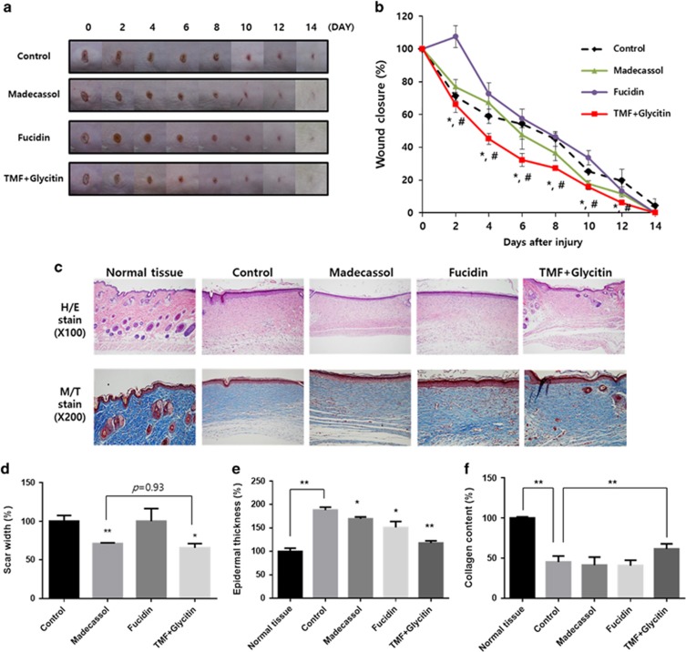
Keratinocyte-fibroblast interactions are critical for skin repair after injury. During the proliferative phase of wound healing, proliferation, migration and differentiation of these cells are the major mechanisms leading to tissue remodeling. We have previously reported that glycitin, a major soy isoflavone, stimulates dermal fibroblast proliferation; and the phytochemical, 4',6,7-trimethoxyisoflavone (TMF), induces migration of HaCaT keratinocyte cells. We therefore investigated whether these compounds display synergistic effects on skin cells during wound healing in vitro and in vivo. Co-treatment with TMF and glycitin synergistically promotes the proliferation and migration of both keratinocytes and dermal fibroblasts, with a 1:1 ratio of these compounds showing the greatest efficacy in our co-culture system. This keratinocyte-fibroblast interaction occurred via the secretion of TGF-β, and the induction of differentiation and proliferation was confirmed in both indirect and direct co-culture assays. In an excisional and burn wound animal model, mice treated with a 1:1 ratio of TMF and glycitin showed faster wound closure, regeneration and scar reduction than even the positive control drug. These data indicate that two isoflavones, TMF and glycitin, act synergistically to promote wound healing and anti-scarring and could potentially be developed together as a bioactive therapeutic for wound treatment.


Conflict of interest statement

The authors declare no conflict of interest.

Figures

Figure 1
Figure 1
The chemical structure of TMF and glycitin. (a) TMF (4′,6,7-trimethoxyisoflavone). (b) Glycitin (4′-hydroxy-6-methoxyisoflavone-7-D-glucoside). TMF, 4′,6,7-trimethoxyisoflavone.
Figure 2
Figure 2
Glycitin and TMF act synergistically to promote the proliferation and migration of HaCaT keratinocytes and human dermal fibroblasts in culture. (a) Proliferation of dermal fibroblasts treated with either glycitin and TMF, mixed in varying ratios or conditioned media from HaCaT keratinocytes treated with same conditions for 24 h, as measured by MTT assay. (b) Scratch wound healing assay with dermal fibroblasts treated as described in a for 24 h. Migration distance was measured using the ImageJ program. (c) Proliferation of HaCaT keratinocytes treated with either glycitin and TMF, mixed in varying ratios or conditioned media from dermal fibroblasts treated with the same conditions for 24 h, as measured by MTT assay. (d) Scratch wound healing assay with HaCaT keratinocytes treated as described in c for 24 h. Migration distance was measured using the ImageJ program. *P<0.05 as compared with control, **P<0.001 as compared with control, G: glycitin, T: TMF. TMF, 4′,6,7-trimethoxyisoflavone.
Figure 3
Figure 3
A 1:1 mixture of glycitin and TMF increases invasive ability of dermal fibroblasts and HeCaT cells in co-culture conditions via induction of a secreted factor. (a, b) Transwell invasion assay with HaCaT keratinocytes treated with G:T=1:1 (10 μM:10 μM) for 48 h; 7 × 10⁴ cells per well were seeded on the insert, and dermal fibroblasts were also seeded on bottom for co-culture. Invasive ability was measured as described in the ‘Materials and methods' section and is quantified in b. (c, d) Transwell invasion assay with dermal fibroblasts treated with G:T=1:1 (10 μM:10 μM) for 48 h; 7 × 10⁴ cells per well were seeded on the insert, and HeCaT keratinocytes were also seeded on bottom for co-culture. Invasive ability was measured as described in the ‘Materials and methods' section and is quantified in d. (e) Diagram illustrating the direct co-culture system utilized for the invasion assays. Arrows indicate HaCaT keratinocytes, and arrowheads indicate dermal fibroblasts. (f) Invasion assay with HaCaT keratinocytes and dermal fibroblasts in direct contact, using HaCaT keratinocytes transfected with a vector expressing the mCherry fluorescent protein. Invasive ability was measured as described in the ‘Materials and methods' section. *P<0.05 as compared with control, **P<0.001 as compared with control, G: glycitin, T: TMF.
Figure 4
Figure 4
Co-treatment with glycitin and TMF promotes differentiation and proliferation in co-culture condition via secretion of TGF-β. (a) HaCaT keratinocyte and dermal fibroblast cells (1 × 10⁵ each) were seeded on 100 mm cell culture dishes and incubated for 24 h. Protein lysates from cells treated with glycitin (G-10 μM and G-20 μM), TMF (T-10 μM and T-20 μM) or G:T=1:1 (10 μM:10 μM) were obtained using RIPA solution, and proteins were resolved by 10% sodium dodecyl sulfate-polyacrylamide gel electrophoresis. Western blot to measure the expression of differentiation and proliferation-related factors was performed, and in all cases, expression levels were normalized to GAPDH. (b) Western blot assay to detect TFG-β expression in conditioned media from co-cultured cells treated with G:T=1:1 (10 μM:10 μM), G:T=1:2 (6.7 μM:13.3 μM) and G:T=2:1 (13.3 μM:6.7 μM) for 24 h. TGF-β expression was measured by comparing with ponceuS. (c) RT-PCR to measure TGF-β transcript levels in co-cultured cells from transwell assays; TGF-β expression levels were normalized to GAPDH. (d) Western blot assay to detect phospho-Smad2 and phosphor-Smad3 in cell lysates from single-cultured and co-cultured cells after 24 h. *P<0.05 as compared with control, **P<0.001 as compared with control, G: glycitin, T: TMF. TMF, 4′,6,7-trimethoxyisoflavone; TGF-β, transforming growth factor-beta.
Figure 5
Figure 5
A 1:1 mixture of glycitin and TMF accelerates wound closure and protects against scar formation in an in vivo excisional wound model. (a, b) Wound closure in our excisional wound model after 2 weeks. Photographs were taken every 2 days, and wounds were measured using the Image J program. ‘Control' indicates the butylene glycol; *P<0.05 as compared with Madecassol, #P<0.001 as compared with Fucidin. (c) On the last day of treatment, skin tissues were isolated, fixed and stained with hematoxylin/eosin and Masson's trichrome. (d) Scar width, (e) epidermal thickness and (f) collagen content were measured 14 days after wounding using the Image J program. TMF, 4′,6,7-trimethoxyisoflavone.
Figure 6
Figure 6
A 1:1 mixture of glycitin and TMF reduces scarring an in vivo burn wound model. (a) Burn wound closure after treatment for 10 days. (b) Wound sites were treated every day for 8 weeks; hair was then removed, and the scars were photographed using a digital camera or (c) the skin testing machine. (d) Scar size was measured 8 weeks after wounding using the Image J program. (e–h) Skin tissues were isolated, fixed and stained with both hematoxylin/eosin and Masson's trichrome. Epidermal thickness and collagen content were measured using the Image J program. (i) Western blot analysis of skin tissue isolated from wound sites 4 weeks and (j) 8 weeks after wounding. *P<0.05 as compared with control, **P<0.001 as compared with control, G: glycitin, T: TMF. TMF, 4′,6,7-trimethoxyisoflavone.
Figure 6
Figure 6
A 1:1 mixture of glycitin and TMF reduces scarring an in vivo burn wound model. (a) Burn wound closure after treatment for 10 days. (b) Wound sites were treated every day for 8 weeks; hair was then removed, and the scars were photographed using a digital camera or (c) the skin testing machine. (d) Scar size was measured 8 weeks after wounding using the Image J program. (e–h) Skin tissues were isolated, fixed and stained with both hematoxylin/eosin and Masson's trichrome. Epidermal thickness and collagen content were measured using the Image J program. (i) Western blot analysis of skin tissue isolated from wound sites 4 weeks and (j) 8 weeks after wounding. *P<0.05 as compared with control, **P<0.001 as compared with control, G: glycitin, T: TMF. TMF, 4′,6,7-trimethoxyisoflavone.

References

    1. Harborne JB, Williams CA. Advances in flavonoid research since 1992. Phytochemistry 2000; 55: 481–504. - PubMed
    1. Pietta PG. Flavonoids as antioxidants. J Nat Prod 2000; 63: 1035–1042. - PubMed
    1. Yao LH, Jiang YM, Shi J, Tomas-Barberan FA, Datta N, Singanusong R et al. Flavonoids in food and their health benefits. Plant Food Hum Nutr 2004; 59: 113–122. - PubMed
    1. Lee CH, Yang L, Xu JZ, Yeung SYV, Huang Y, Chen ZY. Relative antioxidant activity of soybean isoflavone and their glycosides. Food Chem 2005; 90: 735–741.
    1. Song TT, Hendrich S, Murphy PA. Estrogenic activity of glycitein, a soy isoflavone. J Agric Food Chem 1999; 47: 1607–1610. - PubMed

Publication types

LinkOut - more resources