. 2017 Mar 16;7(1):214.
doi: 10.1038/s41598-017-00320-7.

A Biologically-validated HCV E1E2 Heterodimer Structural Model

Affiliations

A Biologically-validated HCV E1E2 Heterodimer Structural Model

Matteo Castelli et al. Sci Rep. .

Abstract

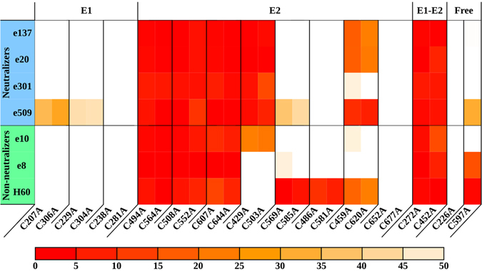
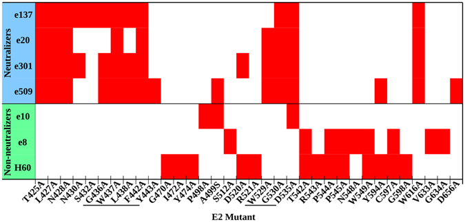
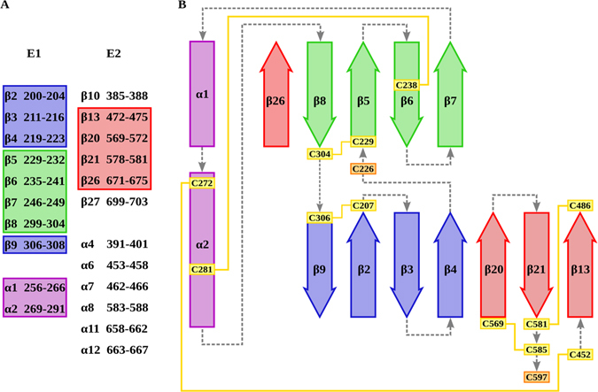
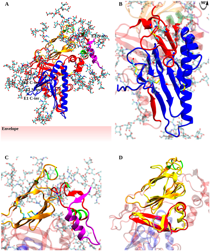
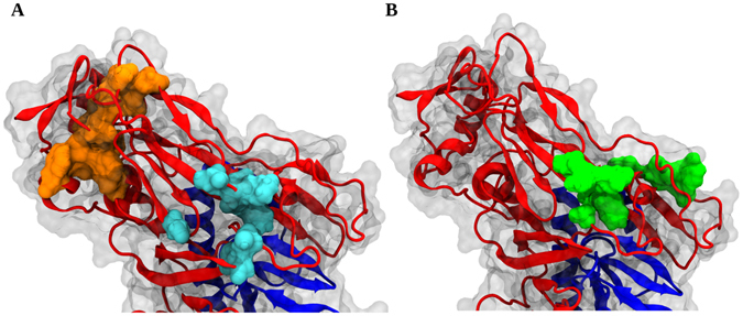
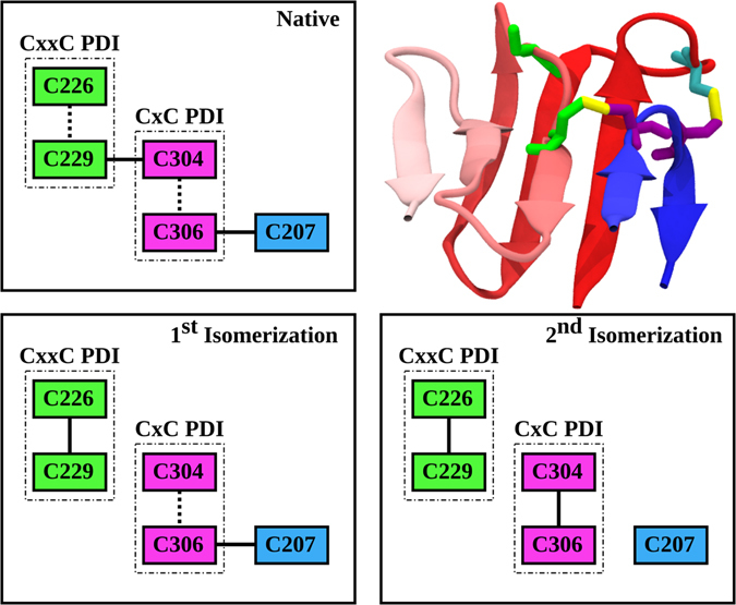
The design of vaccine strategies and the development of drugs targeting the early stages of Hepatitis C virus (HCV) infection are hampered by the lack of structural information about its surface glycoproteins E1 and E2, the two constituents of HCV entry machinery. Despite the recent crystal resolution of limited versions of both proteins in truncated form, a complete picture of the E1E2 complex is still missing. Here we combined deep computational analysis of E1E2 secondary, tertiary and quaternary structure with functional and immunological mutational analysis across E1E2 in order to propose an in silico model for the ectodomain of the E1E2 heterodimer. Our model describes E1-E2 ectodomain dimerization interfaces, provides a structural explanation of E1 and E2 immunogenicity and sheds light on the molecular processes and disulfide bridges isomerization underlying the conformational changes required for fusion. Comprehensive alanine mutational analysis across 553 residues of E1E2 also resulted in identifying the epitope maps of diverse mAbs and the disulfide connectivity underlying E1E2 native conformation. The predicted structure unveils E1 and E2 structures in complex, thus representing a step towards the rational design of immunogens and drugs inhibiting HCV entry.


Conflict of interest statement

The authors declare that they have no competing interests.

Figures

Figure 1
Figure 1
C-to-A reactivity pattern. Antibodies are divided into neutralizers and non-neutralizers. Each cysteine mutant was tested against the mAb panel. Disulfide bridges and free cysteines selected on the basis of their reactivity are reported. Reactivities below 50% compared to WT are depicted as a heatmap spanning from light orange (50%) to red (0%).
Figure 2
Figure 2
Critical residues abrogating mAb binding. Antibodies are divided into neutralizers and non-neutralizers. Epitope residues are marked in red, while residues that are part of the CD81bs are underlined. The threshold below which a decrease in reactivity defines a residue as part of an epitope was determined for each antibody (e137 < 15%, e20 < 20%, e301 < 20%, e509 < 15%, e10 < 30%, e8 < 20%, H60 < 15%).
Figure 3
Figure 3
Secondary and tertiary structure prediction. In (A) all newly identified secondary structures in E1 and E2 are reported. (B) Secondary, tertiary and quaternary arrangement of E1 and E1E2 interfaces. In both panels, previously characterized domains are omitted. Strands and helices, respectively represented as arrows and rectangles, follow the same color scheme in both panels. Successive secondary structure elements are connected by dashed gray lines. Cysteines involved in disulfide bonds are boxed in yellow and connected by yellow lines, free cysteines are boxed in orange.
Figure 4
Figure 4
E1E2 heterodimer model. E1 and E2 structures are represented in cartoon and colored in blue and red, respectively. E2 HVR1, Ig-like domain and CD81bs are colored in purple, orange and green, respectively. Glycosylations are in licorice representation. (A) Overview of the entire, fully-glycosylated structure. (B) Details of E1, E1-E2 interfaces and the protective role of the variable regions HVR2 and IgVR on E1. These domains are depicted in solid colors, while the remaining E2 segments are transparent. (C) Protective role of HVR1 on CD81bs. Residues included in the CD81bs, Ig-like domain and HVR1 are in solid colors, all other regions are transparent. (D) Structural alignment of the E2c structured portions and the E1E2 model. 4MWF structure is aligned to the same regions of the model and colored in solid yellow. Model domains not superimposed to 4MWF are transparent. Glycosylations were removed for clarity.
Figure 5
Figure 5
Epitope mapping on E1E2 model. E1 and E2 are represented in transparent surface and blue and red cartoon, respectively. (A) A non-neutralizing mAb (e8) and a broad neutralizing mAb (e137) epitopes are highlighted in cyan and orange, respectively. (B) H60 epitope is depicted in green.
Figure 6
Figure 6
E1 disulfide isomerization. E1 β-sheets structure in the native conformation is depicted in cartoon and colored according to residue position (red residues are located at the N-terminal, blue residues at the C-terminal) to highlight long-range β-strand coupling. Cysteines are represented in licorice and colored according to the scheme. In the three boxes, cysteine coupling in native conditions and after the first and second disulfide isomerization steps are represented. Continuous lines connecting each cysteine represent disulfide bridges, while dashed lines highlight proximal cysteines in the linear sequence.

References

    1. Kielian M, Chanel-Vos C, Liao M. Alphavirus Entry and Membrane Fusion. Viruses. 2010;2:796–825. doi: 10.3390/v2040796. - DOI - PMC - PubMed
    1. Modis Y. Relating structure to evolution in class II viral membrane fusion proteins. Current opinion in virology. 2014;5:34–41. doi: 10.1016/j.coviro.2014.01.009. - DOI - PMC - PubMed
    1. Lorenz IC, Allison SL, Heinz FX, Helenius A. Folding and dimerization of tick-borne encephalitis virus envelope proteins prM and E in the endoplasmic reticulum. Journal of virology. 2002;76:5480–5491. doi: 10.1128/JVI.76.11.5480-5491.2002. - DOI - PMC - PubMed
    1. El Omari K, Iourin O, Harlos K, Grimes JM, Stuart DI. Structure of a pestivirus envelope glycoprotein E2 clarifies its role in cell entry. Cell reports. 2013;3:30–35. doi: 10.1016/j.celrep.2012.12.001. - DOI - PMC - PubMed
    1. Catanese MT, et al. Ultrastructural analysis of hepatitis C virus particles. Proceedings of the National Academy of Sciences of the United States of America. 2013;110:9505–9510. doi: 10.1073/pnas.1307527110. - DOI - PMC - PubMed

Substances

LinkOut - more resources