Review
. 2017 Mar 2:8:198.
doi: 10.3389/fimmu.2017.00198. eCollection 2017.

Cellular and Molecular Characterization of Microglia: A Unique Immune Cell Population

Affiliations
Review

Cellular and Molecular Characterization of Microglia: A Unique Immune Cell Population

Carole Sousa et al. Front Immunol. .

Abstract

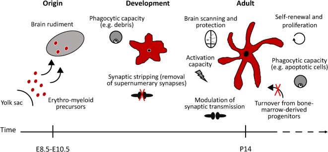
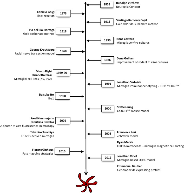
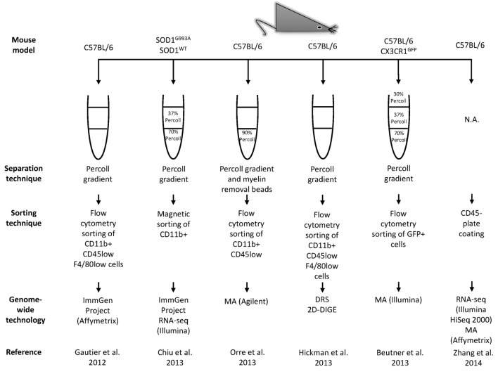
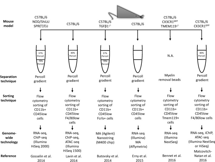
Microglia are essential for the development and function of the adult brain. Microglia arise from erythro-myeloid precursors in the yolk sac and populate the brain rudiment early during development. Unlike monocytes that are constantly renewed from bone marrow hematopoietic stem cells throughout life, resident microglia in the healthy brain persist during adulthood via constant self-renewal. Their ontogeny, together with the absence of turnover from the periphery and the singular environment of the central nervous system, make microglia a unique cell population. Supporting this notion, recent genome-wide transcriptional studies revealed specific gene expression profiles clearly distinct from other brain and peripheral immune cells. Here, we highlight the breakthrough studies that, over the last decades, helped elucidate microglial cell identity, ontogeny, and function. We describe the main techniques that have been used for this task and outline the crucial milestones that have been achieved to reach our actual knowledge of microglia. Furthermore, we give an overview of the "microgliome" that is currently emerging thanks to the constant progress in the modern profiling techniques.

Keywords: Rio Hortega; genome-wide; microglia history; microgliome; technology.


Figures

Figure 1
Figure 1
Schematic representation of microglial functional states in the healthy murine brain. Microglia arise from erythro-myeloid precursors in the embryonic yolk sac and populate the brain rudiment early during development. Microglial cell population is maintained by self-renewal, without the contribution of bone marrow-derived progenitors. In the adult healthy brain, microglia continuously survey the brain and readily react to any potential threat to the CNS homeostasis. Phagocytic microglia can detect and quickly remove damaged or dying neurons, preventing further damage to neighboring cells. During developmental stages, microglia phagocytic capacity is particularly important to prune supernumerary synapses. Microglia has also been suggested to modulate neuronal activity by influencing synapse transmission (synaptic stripping). Under specific conditions, microglia are able to remove dysfunctional synapses by physically interacting with functional neurons.
Figure 2
Figure 2
Timeline of the main techniques and methodologies used in microglia research. Major approaches that have contributed to breakthrough findings to elucidate microglial cells identity, ontogeny, and function.
Figure 3
Figure 3
Continued Workflow illustrating technical and methodological details used in the main genome-wide gene expression profiling studies. The related results are shown in Table 2. MA, microarrays; RNA-seq, RNA-sequencing; DRS, direct RNA sequencing; 2D-DIGE, two-dimensional difference gel electrophoresis; ChIP-seq, chromatin immunoprecipitation sequencing; ATAC-seq, assay for transposase accessible chromatin; iChIP, indexing-first chromatin immunoprecipitation.
Figure 3
Figure 3
Continued Workflow illustrating technical and methodological details used in the main genome-wide gene expression profiling studies. The related results are shown in Table 2. MA, microarrays; RNA-seq, RNA-sequencing; DRS, direct RNA sequencing; 2D-DIGE, two-dimensional difference gel electrophoresis; ChIP-seq, chromatin immunoprecipitation sequencing; ATAC-seq, assay for transposase accessible chromatin; iChIP, indexing-first chromatin immunoprecipitation.

References

    1. Ransohoff RM, Engelhardt B. The anatomical and cellular basis of immune surveillance in the central nervous system. Nat Rev Immunol (2012) 12:623–35.10.1038/nri3265 - DOI - PubMed
    1. Kipnis J. Multifaceted interactions between adaptive immunity and the central nervous system. Science (2016) 353:766–71.10.1126/science.aag2638 - DOI - PMC - PubMed
    1. Louveau A, Da Mesquita S, Kipnis J. Lymphatics in neurological disorders: a neuro-lympho-vascular component of multiple sclerosis and Alzheimer’s disease? Neuron (2016) 91:957–73.10.1016/j.neuron.2016.08.027 - DOI - PMC - PubMed
    1. Perry VH, Nicoll JA, Holmes C. Microglia in neurodegenerative disease. Nat Rev Neurol (2010) 6:193–201.10.1038/nrneurol.2010.17 - DOI - PubMed
    1. Gordon S, Pluddemann A, Martinez Estrada F. Macrophage heterogeneity in tissues: phenotypic diversity and functions. Immunol Rev (2014) 262:36–55.10.1111/imr.12223 - DOI - PMC - PubMed