. 2017 Jul;101(1):92-101.
doi: 10.1007/s00223-017-0259-2. Epub 2017 Mar 16.

Bone Alkaline Phosphatase and Tartrate-Resistant Acid Phosphatase: Potential Co-regulators of Bone Mineralization

Affiliations

Bone Alkaline Phosphatase and Tartrate-Resistant Acid Phosphatase: Potential Co-regulators of Bone Mineralization

Cecilia Halling Linder et al. Calcif Tissue Int. 2017 Jul.

Abstract

Phosphorylated osteopontin (OPN) inhibits hydroxyapatite crystal formation and growth, and bone alkaline phosphatase (BALP) promotes extracellular mineralization via the release of inorganic phosphate from the mineralization inhibitor inorganic pyrophosphate (PPi). Tartrate-resistant acid phosphatase (TRAP), produced by osteoclasts, osteoblasts, and osteocytes, exhibits potent phosphatase activity towards OPN; however, its potential capacity as a regulator of mineralization has not previously been addressed. We compared the efficiency of BALP and TRAP towards the endogenous substrates for BALP, i.e., PPi and pyridoxal 5'-phosphate (PLP), and their impact on mineralization in vitro via dephosphorylation of bovine milk OPN. TRAP showed higher phosphatase activity towards phosphorylated OPN and PPi compared to BALP, whereas the activity of TRAP and BALP towards PLP was comparable. Bovine milk OPN could be completely dephosphorylated by TRAP, liberating all its 28 phosphates, whereas BALP dephosphorylated at most 10 phosphates. OPN, dephosphorylated by either BALP or TRAP, showed a partially or completely attenuated phosphorylation-dependent inhibitory capacity, respectively, compared to native OPN on the formation of mineralized nodules. Thus, there are phosphorylations in OPN important for inhibition of mineralization that are removed by TRAP but not by BALP. In conclusion, our data indicate that both BALP and TRAP can alleviate the inhibitory effect of OPN on mineralization, suggesting a potential role for TRAP in skeletal mineralization. Further studies are warranted to explore the possible physiological relevance of TRAP in bone mineralization.

Keywords: Bone; Dephosphorylation; Hydroxyapatite; Inorganic pyrophosphate; Mineralization; Osteopontin.


Conflict of interest statement

Conflict of interest

The authors Cecilia Halling Linder, Barbro Ek-Rylander, Michael Krumpel, Maria Norgård, Sonoko Narisawa, José Luis Millán, Göran Andersson, and Per Magnusson declare that they have no conflict of interest.

Human and Animal Rights and Informed Consent

This article does not contain any studies with human or animal subjects performed by any of the authors.

Figures

Fig. 1
Fig. 1
Dephosphorylation of OPN by TRAP, BALP, and IALP. OPN was completely dephosphorylated (28 free phosphates liberated) after 24-h incubation with 5 mU of TRAP (filled triangle). During the same time period, 20 mU of BALP (filled square) cleaved off 10 free phosphates and 20 mU IALP (open square) liberated 20 free phosphates from OPN. All experiments were run in triplicate. Statistical comparisons were made between TRAP, BALP, and IALP at each time point (NS not significant)
Fig. 2
Fig. 2
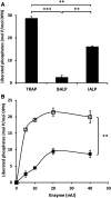
A Differences between TRAP, BALP, and IALP regarding the dephosphorylation efficiency on OPN. These results demonstrate the amount of liberated free phosphate cleaved off from OPN after 24-h incubation with 5 mU of TRAP, BALP, and IALP. B Maximum amount of phosphate liberated from the OPN molecule after 24 h of incubation with different concentrations of BALP (filled square) and IALP (open square). Statistical comparisons were made between the maximum values of liberated free phosphate for each enzyme after 24-h incubation of each enzyme. Results are presented as mean ± SD of three independent experiments. ** P < 0.01, *** P < 0.001
Fig. 3
Fig. 3
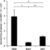
Dephosphorylation of OPN with TRAP, BALP, and IALP with respect to liberated free phosphate per mol OPN calculated per hour and mU enzyme. * P < 0.05, ** P < 0.01
Fig. 4
Fig. 4
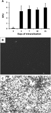
A Osteoblast-like SaOS-2 cells cultured for 3, 5, 7, 10, and 21 days after initiation of mineralization (without OPN). Cells were stained with the OsteoImage Mineralization Assay, which specifically detects the HA portion of bone-like nodules deposited by cells. Fluorescence was measured in relative fluorescence units (RFU) and is proportional to the amount of HA present in the culture. Results are presented as mean ± SD of eight samples. B Image of cells cultured for 10 days without mineralization medium. C Image of cells cultured for 10 days with mineralization medium (without OPN)
Fig. 5
Fig. 5
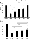
Amount of mineralization in osteoblast-like SaOS-2 cells 5 days after initiation of mineralization. Cells grown with mineralization medium but without OPN were defined as controls and considered to be fully mineralized, and set to 100% relative fluorescence units (RFU). The other bars are expressed as percentages of 100% RFU. +OPN indicates the addition of OPN (fully phosphorylated), and −2P, −5P, −10P, and −28P indicate the number of phosphates cleaved off (dephosphorylated OPN). OPN, fully phosphorylated and dephosphorylated, was added at a final concentration of 0.1 µg/mL. Results are presented as mean ± SD of three independent experiments with eight samples in each experiment. A OPN fully phosphorylated and dephosphorylated by TRAP. B OPN fully phosphorylated and dephosphorylated by BALP. * P < 0.05, ** P < 0.01, *** P < 0.005, ns not significant

Similar articles

Cited by

References

    1. McNally EA, Schwarcz HP, Botton GA, Arsenault AL. A model for the ultrastructure of bone based on electron microscopy of ion-milled sections. PLoS ONE. 2012;7:e29258. doi: 10.1371/journal.pone.0029258. - DOI - PMC - PubMed
    1. Mahamid J, Addadi L, Weiner S. Crystallization pathways in bone. Cells Tissues Organs. 2011;194:92–97. doi: 10.1159/000324229. - DOI - PubMed
    1. Millán JL. The role of phosphatases in the initiation of skeletal mineralization. Calcif Tissue Int. 2013;93:299–306. doi: 10.1007/s00223-012-9672-8. - DOI - PMC - PubMed
    1. Hunter GK. Role of osteopontin in modulation of hydroxyapatite formation. Calcif Tissue Int. 2013;93:348–354. doi: 10.1007/s00223-013-9698-6. - DOI - PubMed
    1. Cui L, Houston DA, Farquharson C, MacRae VE. Characterisation of matrix vesicles in skeletal and soft tissue mineralisation. Bone. 2016;87:147–158. doi: 10.1016/j.bone.2016.04.007. - DOI - PubMed

Publication types