

Morphea and Eosinophilic Fasciitis: An Update
- PMID: 28303481
- PMCID: PMC5506513
- DOI: 10.1007/s40257-017-0269-x
Morphea and Eosinophilic Fasciitis: An Update
Abstract
Morphea, also known as localized scleroderma, encompasses a group of idiopathic sclerotic skin diseases. The spectrum ranges from relatively mild phenotypes, which generally cause few problems besides local discomfort and visible disfigurement, to subtypes with severe complications such as joint contractures and limb length discrepancies. Eosinophilic fasciitis (EF, Shulman syndrome) is often regarded as belonging to the severe end of the morphea spectrum. The exact driving mechanisms behind morphea and EF pathogenesis remain to be elucidated. However, extensive extracellular matrix formation and autoimmune dysfunction are thought to be key pathogenic processes. Likewise, these processes are considered essential in systemic sclerosis (SSc) pathogenesis. In addition, similarities in clinical presentation between morphea and SSc have led to many theories about their relatedness. Importantly, morphea may be differentiated from SSc based on absence of sclerodactyly, Raynaud's phenomenon, and nailfold capillary changes. The diagnosis of morphea is often based on characteristic clinical findings. Histopathological evaluation of skin biopsies and laboratory tests are not necessary in the majority of morphea cases. However, full-thickness skin biopsies, containing fascia and muscle tissue, are required for the diagnosis of EF. Monitoring of disease activity and damage, especially of subcutaneous involvement, is one of the most challenging aspects of morphea care. Therefore, data harmonization is crucial for optimizing standard care and for comparability of study results. Recently, the localized scleroderma cutaneous assessment tool (LoSCAT) has been developed and validated for morphea. The LoSCAT is currently the most widely reported outcome measure for morphea. Care providers should take disease subtype, degree of activity, depth of involvement, and quality-of-life impairments into account when initiating treatment. In most patients with circumscribed superficial subtypes, treatment with topical therapies suffices. In more widespread disease, UVA1 phototherapy or systemic treatment with methotrexate (MTX), with or without a systemic corticosteroid combination, should be initiated. Disappointingly, few alternatives for MTX have been described and additional research is still needed to optimize treatment for these debilitating conditions. In this review, we present a state-of-the-art flow chart that guides care providers in the treatment of morphea and EF.
Conflict of interest statement
Funding
No funding was received for the preparation of this review.
Conflict of interest
Jorre S. Mertens, Marieke M.B. Seyger, Rogier M. Thurlings, Timothy R.D.J. Radstake, and Elke M.G.J. de Jong have no conflicts of interest.
Figures


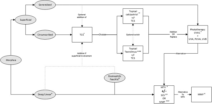
 Methotrexate (MTX): adult starting dose 15 mg/week, max dose 25 mg/week; pediatric starting dose 15 mg/m2, max dose 25 mg. Folic acid supplementation: 0.4–1 mg/day or 5–10 mg/week.
  Systemic corticosteroids (SCS): adult starting dose 0.5–1 mg/kg/day (max 60 mg) during a max of 3 months followed by tapering; pediatric dose: 1–2 mg/kg/day, max dose 60mg/day, followed by tapering.
  Intravenous methylprednisolone (IVMP): adult dose 1000 mg/day for 3 days/month for 3–6 months, possibly followed by oral SCS. Pediatric dose 30 mg/kg/day for 3 days/month for 3 months, possibly followed by oral SCS. α Mycophenolate Mofetil (MMF, alternative to MTX): adult dose 1000 mg twice daily. Pediatric dose 600–1200 mg/m2/day twice daily. A Deep/linear subtypes: treatment with MTX monotherapy. Addition of SCS or IVMP in case of rapidly progressive disease or in the presence of (looming) contractures. B Eosinophilic Fasciitis: standard induction treatment with oral SCS or IVMP in combination with MTX . PUVA psoralen plus broadband UVA, UV ultraviolet
References
-
- Christen-Zaech S, Hakim MD, Afsar FS, Paller AS. Pediatric morphea (localized scleroderma): review of 136 patients. J Am Acad Dermatol. 2008;59(3):385–396. - PubMed
-
- Nelson AM. Localized scleroderma including morphea, linear scleroderma, and eosinophilic fasciitis. Curr Probl Pediatr. 1996;26(9):318–324. - PubMed
-
- Mazori DR, Wright NA, Patel M, Liu SW, Ramachandran SM, Franks AG, Jr, et al. Characteristics and treatment of adult-onset linear morphea: a retrospective cohort study of 61 patients at 3 tertiary care centers. J Am Acad Dermatol. 2016;74(3):577–579. - PubMed
-
- Zulian F, Vallongo C, Woo P, Russo R, Ruperto N, Harper J, et al. Localized scleroderma in childhood is not just a skin disease. Arthritis Rheum. 2005;52(9):2873–2881. - PubMed
Publication types
MeSH terms
Substances
Supplementary concepts
LinkOut - more resources
Full Text Sources
Other Literature Sources
Medical
