. 2017 Mar;24(2):10.1111/xen.12293.
doi: 10.1111/xen.12293. Epub 2017 Mar 17.

Immunological and physiological observations in baboons with life-supporting genetically engineered pig kidney grafts

Affiliations

Immunological and physiological observations in baboons with life-supporting genetically engineered pig kidney grafts

Hayato Iwase et al. Xenotransplantation. 2017 Mar.

Abstract

Background: Genetically engineered pigs could provide a source of kidneys for clinical transplantation. The two longest kidney graft survivals reported to date have been 136 and 310 days, but graft survival >30 days has been unusual until recently.

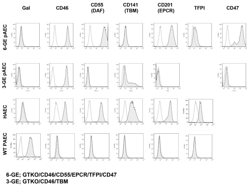
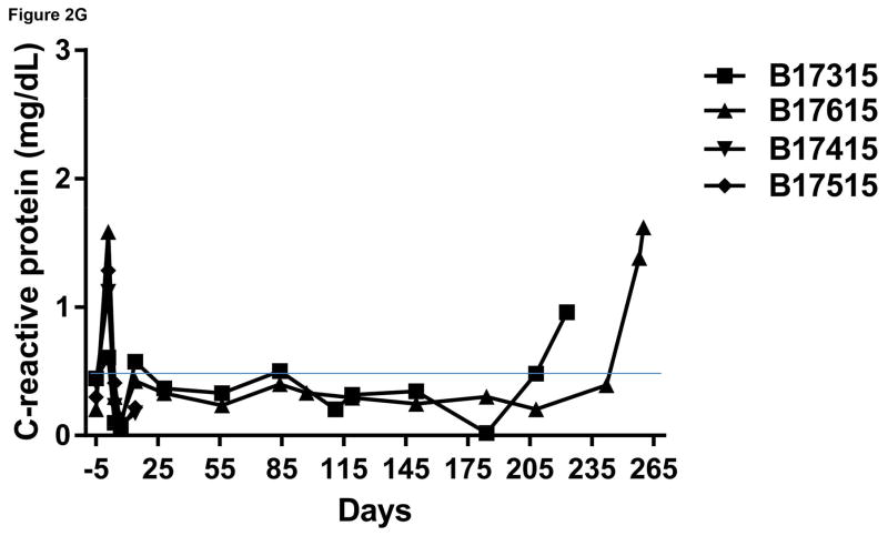
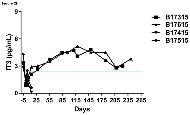
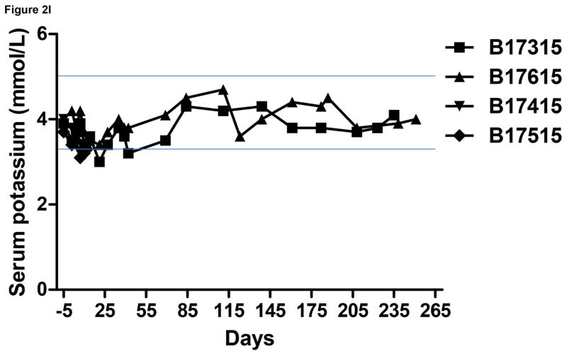
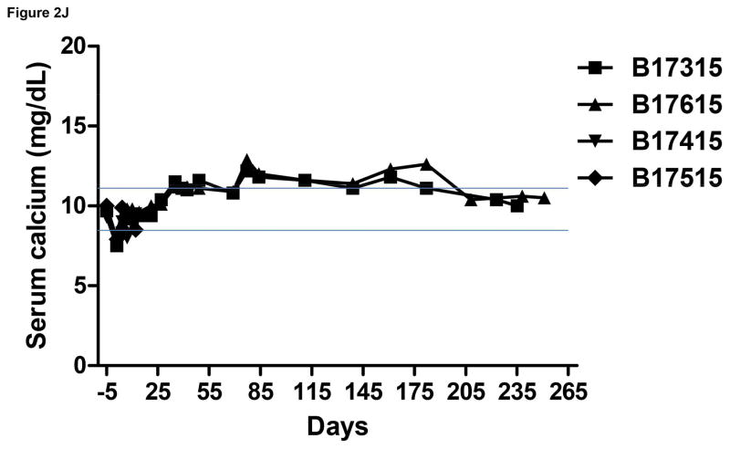
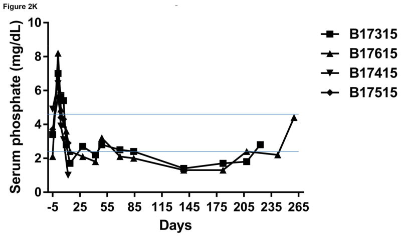
Methods: Donor pigs (n=4) were on an α1,3-galactosyltransferase gene-knockout (GTKO)/human complement regulatory protein (CD46) background (GTKO/CD46). In addition, the pigs were transgenic for at least one human coagulation regulatory protein. Two baboons received a kidney from a six-gene pig (GroupA) and two from a three-gene pig (GroupB). Immunosuppressive therapy was identical in all four cases and consisted of anti-thymoglobulin (ATG)+anti-CD20mAb (induction) and anti-CD40mAb+rapamycin+corticosteroids (maintenance). Anti-TNF-α and anti-IL-6R mAbs were administered to reduce the inflammatory response. Baboons were followed by clinical/laboratory monitoring of immune/coagulation/inflammatory/physiological parameters. At biopsy or euthanasia, the grafts were examined by microscopy.

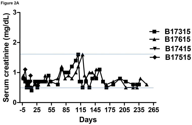
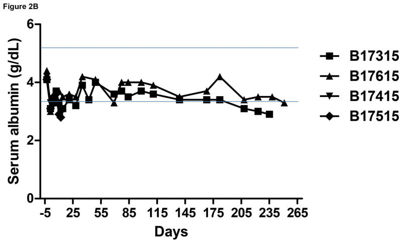
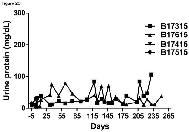
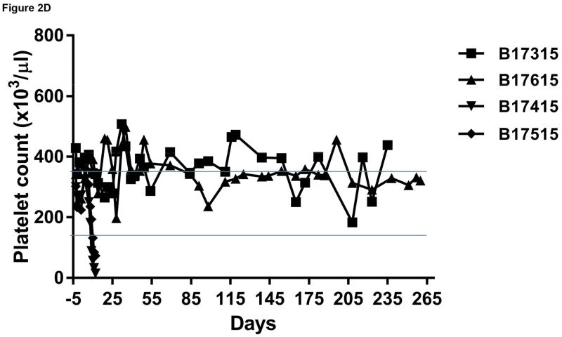
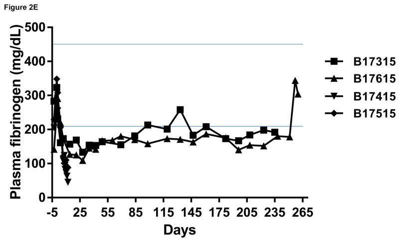
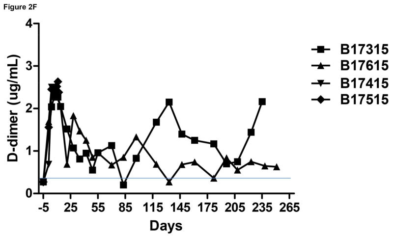
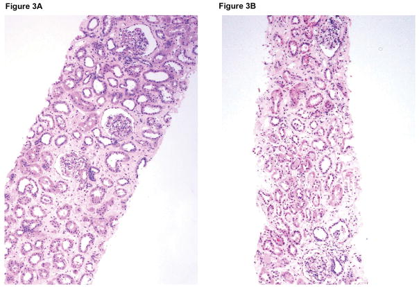
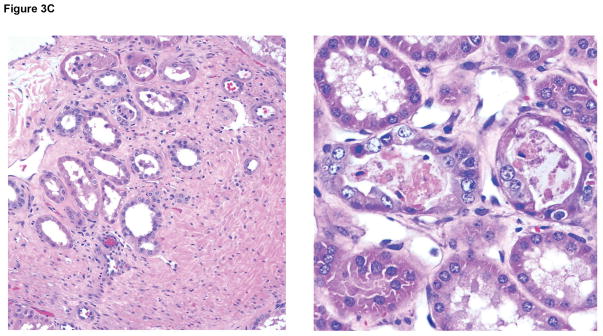
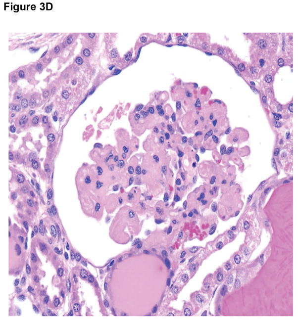
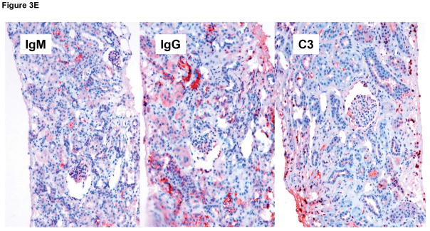
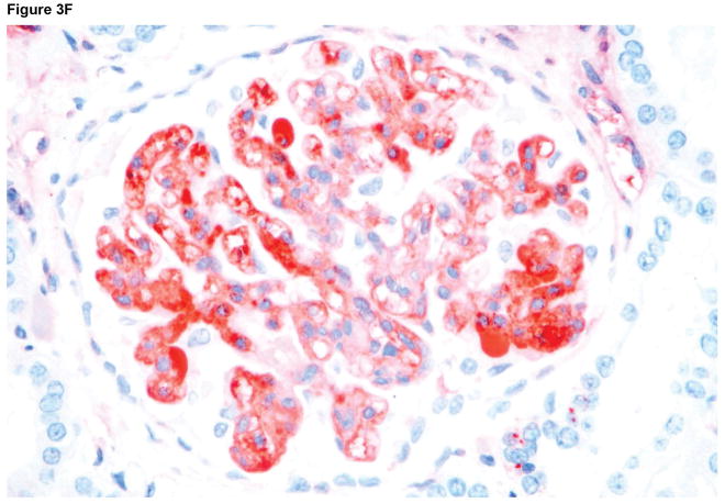
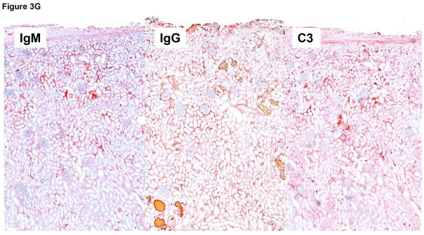
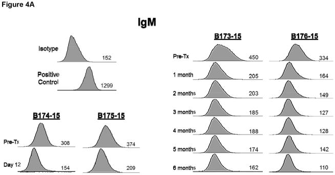
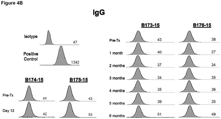
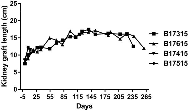
Results: The two GroupA baboons remained healthy with normal renal function >7 and >8 months, respectively, but then developed infectious complications. However, no features of a consumptive coagulopathy, eg, thrombocytopenia and reduction of fibrinogen, or of a protein-losing nephropathy were observed. There was no evidence of an elicited anti-pig antibody response, and histology of biopsies taken at approximately 4, 6, and 7 months and at necropsy showed no significant abnormalities. In contrast, both GroupB baboons developed features of a consumptive coagulopathy and required euthanasia on day 12.

Conclusions: The combination of (i) a graft from a specific six-gene genetically modified pig, (ii) an effective immunosuppressive regimen, and (iii) anti-inflammatory therapy prevented immune injury, a protein-losing nephropathy, and coagulation dysfunction for >7 months. Although the number of experiments is very limited, our impression is that expression of human endothelial protein C receptor (±CD55) in the graft is important if coagulation dysregulation is to be avoided.

Keywords: anti-IL-6R antagonist; costimulation blockade; genetically engineered; kidney; pig; xenotransplantation.


Conflict of interest statement

DISCLOSURE OF CONFLICT OF INTEREST

David Ayares and Carol Phelps are employees of Revivicor, Inc. No other author has a conflict of interest.

Figures

Figure 1
Figure 1
Expression of human transgenes on donor pAECs by flow cytometry. Gal expression was absent and expression of CD46 was high in both donor pigs. The expression of CD55, EPCR, and CD47 was high on pAECs in the 6-gene donor pig, with some expression of TFPI. In the 3-gene pig, there was moderate expression of thrombomodulin.
Figure 2
Figure 2
Monitoring of recipient baboon throughout the course of the experiment. (A) serum creatinine; (B) serum albumin; (C) urinary protein; (D) platelet counts; (E) plasma fibrinogen; (F) D-dimer; (G) C-reactive protein; (H) free triiodothyronine (fT3); (I) serum potassium; (J) serum calcium; (K) serum phosphate. The horizontal lines indicate either the upper and lower limits (A, B, D, E, H, I, J, K), or upper limit (F, G), of the equivalent parameters measured in healthy humans.
Figure 2
Figure 2
Monitoring of recipient baboon throughout the course of the experiment. (A) serum creatinine; (B) serum albumin; (C) urinary protein; (D) platelet counts; (E) plasma fibrinogen; (F) D-dimer; (G) C-reactive protein; (H) free triiodothyronine (fT3); (I) serum potassium; (J) serum calcium; (K) serum phosphate. The horizontal lines indicate either the upper and lower limits (A, B, D, E, H, I, J, K), or upper limit (F, G), of the equivalent parameters measured in healthy humans.
Figure 3
Figure 3
Microscopic appearances of the pig kidney grafts at biopsy at 4 and 7 (A) months (B17315) and at 6 (B) months (B17615) showed no significant abnormalities. At necropsy (at 237 days), the pig kidney in B17615 (C) showed some interstitial fibrosis (left) and mild, patchy acute tubular epithelial cell degeneration in some proximal tubules (right), believed to be secondary to the prolonged hypoxia experienced as a result of the pneumocystis pneumonia. The pig kidney grafts in Group B recipients (D) showed widespread focal hemorrhage and features of thrombotic microangiopathy (multiple occluding thrombi within glomerular capillary loops). Immunohistochemistry staining of the pig kidney grafts at biopsy at 7 months (B17315) and at 6 months (B17615) in Group A (E), demonstrated non-specific deposition (i.e., not localized to the vascular endothelium) of IgM (left), IgG (center), and complement (C3) (right), suggesting no elicited antibody response. In B17315 at euthanasia (day 237) there was more specific staining for IgM in the glomeruli (F), but no specific staining for IgG or complement (not shown). In Group B at necropsy (G), there was non-specific staining for IgM (left), IgG (center), and complement (right).
Figure 3
Figure 3
Microscopic appearances of the pig kidney grafts at biopsy at 4 and 7 (A) months (B17315) and at 6 (B) months (B17615) showed no significant abnormalities. At necropsy (at 237 days), the pig kidney in B17615 (C) showed some interstitial fibrosis (left) and mild, patchy acute tubular epithelial cell degeneration in some proximal tubules (right), believed to be secondary to the prolonged hypoxia experienced as a result of the pneumocystis pneumonia. The pig kidney grafts in Group B recipients (D) showed widespread focal hemorrhage and features of thrombotic microangiopathy (multiple occluding thrombi within glomerular capillary loops). Immunohistochemistry staining of the pig kidney grafts at biopsy at 7 months (B17315) and at 6 months (B17615) in Group A (E), demonstrated non-specific deposition (i.e., not localized to the vascular endothelium) of IgM (left), IgG (center), and complement (C3) (right), suggesting no elicited antibody response. In B17315 at euthanasia (day 237) there was more specific staining for IgM in the glomeruli (F), but no specific staining for IgG or complement (not shown). In Group B at necropsy (G), there was non-specific staining for IgM (left), IgG (center), and complement (right).
Figure 4
Figure 4
Anti-pig nonGal IgM (A) and IgG (B) levels in the recipient baboons (determined by flow cytometry) throughout the post-transplant course.
Figure 4
Figure 4
Anti-pig nonGal IgM (A) and IgG (B) levels in the recipient baboons (determined by flow cytometry) throughout the post-transplant course.
Figure 5
Figure 5
Increases in the lengths of the two Group A pig kidneys determined by ultrasound or direct measurement (at operation).

Similar articles

Cited by

References

    1. Cooper DK, Satyananda V, Ekser B, et al. Progress in pig-to-nonhuman primate transplantation models (1998–2013): a comprehensive review of the literature. Xenotransplantation. 2014;21:397–419. - PMC - PubMed
    1. Lambrigts D, Sachs DH, Cooper DK. Discordant organ xenotransplantation in primates: world experience and current status. Transplantation. 1998;66:547–561. - PubMed
    1. Iwase H, Kobayashi T. Current status of pig kidney xenotransplantation. Int J Surg. 2015;23:229–233. - PMC - PubMed
    1. Baldan N, Rigotti P, Calabrese F, et al. Ureteral stenosis in HDAF pig-to-primate renal xenotransplantation: a phenomenon related to immunological events? Am J Transplant. 2004;4:475–481. - PubMed
    1. Higginbotham L, Mathews D, Breeden CA, et al. Pre-transplant antibody screening and anti-CD154 costimulation blockade promote long-term xenograft survival in a pig-to-primate kidney transplant model. Xenotransplantation. 2015;22:221–230. - PMC - PubMed

Publication types

Substances