. 2017 Mar 17:7:44744.
doi: 10.1038/srep44744.

E6/E7-P53-POU2F1-CTHRC1 axis promotes cervical cancer metastasis and activates Wnt/PCP pathway

Affiliations

E6/E7-P53-POU2F1-CTHRC1 axis promotes cervical cancer metastasis and activates Wnt/PCP pathway

Rong Zhang et al. Sci Rep. .

Abstract

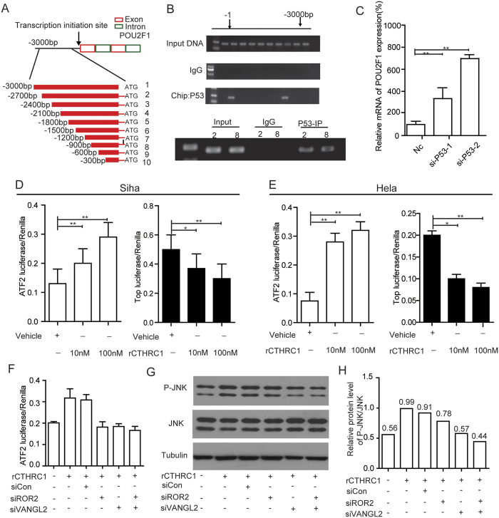
Cervical cancer is an infectious cancer and the most common gynecologic cancer worldwide. E6/E7, the early genes of the high-risk mucosal human papillomavirus type, play key roles in the carcinogenic process of cervical cancer. However, little was known about its roles in modulating tumor microenvironment, particular extracellular matrix (ECM). In this study, we found that E6/E7 could regulate multiple ECM proteins, especially collagen triple helix repeat containing 1 (CTHRC1). CTHRC1 is highly expressed in cervical cancer tissue and serum and closely correlated with clinicopathological parameters. CTHRC1 promotes cervical cancer cell migration and invasion in vitro and metastasis in vivo. E6/E7 regulates the expression of CTHRC1 in cervical cancer by E6/E7-p53-POU2F1 (POU class 2 homeobox 1) axis. Futhermore, CTHRC1 activates Wnt/PCP signaling pathway. Take together, E6/E7-p53-POU2F1-CTHRC1 axis promotes cervical cancer cell invasion and metastasis and may act as a potential therapeutic target for interventions against cervical cancer invasion and metastasis.


Conflict of interest statement

The authors declare no competing financial interests.

Figures

Figure 1
Figure 1. Extracellular matrix expression profile is altered by silencing of E6/E7.
(A) 21 genes that encode extracellular secreted proteins were significantly down-regulated in Caski and Ms751 cells with silencing of E6/E7 as compared with control cells (fold change >3 and P < 0.05). (B) Shows a schematic diagram of down-regulated extracellular secreted proteins. (C) The expression of CTHRC1 in Caski cells with silencing of E6/E7, detected by RT-PCR and normalized with 18S expression. (D) The expression of CTHRC1 in Ms751cells with silencing of E6/E7, detected by RT-PCR and normalized with 18S expression.
Figure 2
Figure 2. CTHRC1 expression was significantly up-regulated in cervical cancers.
(A) The mRNA expression of CTHRC1 is upregulated in tumor tissues compared with the normal tissues revealed using the TCGA dataset. (B) The mRNA expression of CTHRC1 is upregulated in three matched tumor and non-tumor tissue revealed using the TCGA dataset. (C) CTHRC1 expression in the normal and tumor tissues revealed by the GSE31056 dataset. (D) The mRNA expression of CTHRC1 was analyzed in different cancers in Oncomine datasete about 42 women. (E) The copy number of CTHRC1 expression in different tissues was analyzed in TCGA dataset about 200 women.
Figure 3
Figure 3. Immunohistochemical analysis of CTHRC1 in cervical cancer tissues.
(A) Representative photographs of the CTHRC1 immunoreactivity in Normal, Adenocarcinoma, and Squamous cancer tissues. (B) Comparisons of CTHRC1 expression in tissues revealed by IHC analysis in Normal, Adenocarcinoma, and Squamous cancer tissues. (C) Representative photographs of the CTHRC1 staining in grade 1, 2,3 squamous cervical carcinoma tissues. (D) Comparisons of CTHRC1 expression in tissues revealed by IHC analysis grade 1, 2,3 squamous cervical carcinoma tissues. (E) Representative photographs of the CTHRC1 staining in Non-metastasis and metastasis of lymph gland tissues. (F) Comparisons of CTHRC1 expression in tissues revealed by IHC analysis Non-metastasis metastasis of lymph gland tissues. The positive staining of CTHRC1 is shown in brown color, and the cell nuclei were counterstained with hematoxylin. Scale bars, 10 μm. Original magnification: 200 × . Data are means ± SD. *P < 0.05, **P < 0.01.
Figure 4
Figure 4. CTHRC1 have no effect on cells proliferation in vitro and tumor growth in vivo.
(A and B) The expression of CTHRC1 in cell lysates from Caski, Ms751, Siha and Hela cells was detected by RT-PCR and normalized with 18S expression, western blot and normalized with β-actin expression. (C and D) The cell proliferation of Nc and sh-1, sh-2 groups in Caski (C) and Ms751 (D) cells were determined by CCK8 assay at 0, 24, 48, 72, 96 h, respectively. Values are means ± SD, n = 5. (E and F) The cell proliferation of control and Lenti-CTHRC1 in Siha and Hela cells was determined by CCK8 assay at 0, 24, 48, 72, 96 h, respectively. Values are means ± SD, n = 5. (G) Morphologic characteristics of tumors from mice inoculated with Siha/Control and Siha/Lenti-CTHRC1 cells. (H and I) Tumor volumes and tumor weights of Nc and Lenti-CTHRC1 groups from G. n = 6.
Figure 5
Figure 5. Silencing of CTHRC1 suppresses cervical cancer migration and invasion in vitro.
(A and B) Representative wound healing images of Caski (A) and Ms751 (B) at 0 and 48 h, respectively. The black line outlined the cell boundary. (C) Quantification of wound healing rates was analyzed in Caski and Ms751 cells respectively. Data are means ± SD of the wound area relative to the Nc group, n = 3. (D and E) Representative migration images of CTHRC1 silenced and Nc cells. (F) Quantification of migration rates was analyzed in Caski and Ms751cells respectively. (G and H) Representative invasion images of CTHRC1 silenced and Nc cells. (I) Quantification of invasion rates was analyzed in cells. Original magnification: 200 × . Quantifications of cells on the lower surface of the membrane were performed with three randomly selected fields. Data are means ± SD. *P < 0.05, **P < 0.01.
Figure 6
Figure 6. CTHRC1 can promote cervical cancer migration and invasion in vitro.
(A and B) Representative wound healing images of Siha (A) and Hela (B) at 0 and 48 h, respectively. The black line outlined the cell boundary. (C) Quantification of wound healing rates was analyzed in Siha and Hela cells respectively. Data are means ± SD of the wound area relative to the control group, n = 3. (D) Quantification of migrated rates was analyzed in Siha and Hela cells respectively. (E) Quantification of invaded rates was analyzed in Siha and Hela cells respectively. (F) Statistical analysis of the cell migrated stimulated by rCTHRC1 protein. (G) Pulmonary metastases were detected by H&E staining. (H) Statistical analysis of numbers of pulmonary metastatic nodules, n = 3. Original magnification: 200 × . Quantification of cells on the lower surface of the membrane were performed with three randomly selected fields and shown on the right panels. Data are means ± SD. *P < 0.05, **P < 0.01.
Figure 7
Figure 7. POU2F1 can regulate the exprssion of CTHRC1.
(A) Seven transcription factors were significantly down-regulated in Caski and Ms751 cells with silencing of E6/E7 as compared with control cells (fold change > 3 and P < 0.05). (B) Silencing of E6/E7 in Caski significantly decreased POU2F1 expression, detected by RT-PCR and normalized with 18S expression. (C) Transcription factor POU2F1 maybe the target gene combined the transcription profiling microarray and online tools. (D) Schematic structure of CTHRC1 promoter. The red and green rectangles indicate exon and intron of CTHRC1 respectively. The zone between black lines represent the primers used in ChIP analysis. (E) A ChIP assay was performed using chromatin from Caski cells. (F) The expression of CTHRC1 was decreased with silencing of POU2F1 in Caski cells, detected by RT-PCR and normalized with 18S expression.
Figure 8
Figure 8. CTHRC1 activates Wnt/PCP signaling pathway.
(A) Schematic structure of POU2F1 promoter. The red and green rectangles indicate exon and intron of POU2F1 respectively. The zone between black lines represent the primers used in ChIP analysis. (B) A ChIP assay was performed using chromatin from Caski cells. (C) Silencing of P53 in Caski decreased POU2F1 expression, detected by RT-PCR and normalized with 18S expression. (D and E) Dual-luciferase reporter assay showed that rCthrc1 protein inhibited Wnt/β-catenin signaling and noncanonical Wnt/PCP was activated in Siha and Hela cells by rCTHRC1 protein in a dose-dependent manner. (F) Dual-luciferase reporter assay showed the promotive effect of rCTHRC1 protein on Wnt/PCP signaling was almost blocked after sliencing of ROR2, VANGL2 or ROR2 + VANGL2, respectively. (G) Western blot analysis of phosphorylation level of JNK after rCTHRC1 (100 nM) treatment in Hela cells. Treatment of HeLa cells with rCTHRC1 could promote the phosphorylation of JNK, while sliencing of ROR2, VANGL2 or ROR2 + VANGL2, respectively, almost fully reversed this phenomenon. (H) Quantitative analysis of grey value for phospho-JNK/total JNK ratio using ImageJ software.
Figure 9
Figure 9. CTHRC1 is highly expressed in the serum of cervical cancer patients.
(A) Comparisons of CTHRC1 concentration in serum of Cervical intraepithelial neoplasia (CIN), Normal, cervical cancer patients. (B) ROC curves of CTHRC1, SCC-Ag and combined. The results shown are mean ± SD of relative firefly/Renilla ratio.

Similar articles

Cited by

References

    1. Clifford G. M. et al.. Worldwide distribution of human papillomavirus types in cytologically normal women in the International Agency for Research on Cancer HPV prevalence surveys: a pooled analysis. Lancet 366, 991–998, doi: 10.1016/s0140-6736(05)67069-9 (2005). - DOI - PubMed
    1. Silvia de Sanjose W. G. V. Q., Laia Alemany Daan T Geraets, Jo Ellen Klaustermeier. et al.. Human papillomavirus genotype attribution in invasive cervical cancer: a retrospective cross-sectional worldwide study. Lancet Oncology 11, 1048–1056, doi: 10.1016/s1470-2045(10)70230-8 (2010). - DOI - PubMed
    1. Ghittoni R. et al.. The biological properties of E6 and E7 oncoproteins from human papillomaviruses. Virus Genes 40, 1–13, doi: 10.1007/s11262-009-0412-8 (2010). - DOI - PubMed
    1. Gribben J R. A., Gascoyne R. & Lenz G. Targeting the microenvironment. Leuk Lymphoma 51, Suppl 1, 34–40, doi: 10.3109/10428194.2010.500072. (2010). - DOI - PubMed
    1. Xu Y., Xia X. & Pan H. Active autophagy in the tumor microenvironment: A novel mechanism for cancer metastasis. Oncol Lett 5, 411–416, doi: 10.3892/ol.2012.1015 (2013). - DOI - PMC - PubMed

Publication types

MeSH terms

Substances