Review
. 2017 May 15;595(10):3063-3075.
doi: 10.1113/JP272844. Epub 2017 Apr 21.

Molecular mechanisms of tumour invasion: regulation by calcium signals

Affiliations
Review

Molecular mechanisms of tumour invasion: regulation by calcium signals

Oksana Iamshanova et al. J Physiol. .

Abstract

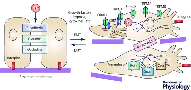
Intracellular calcium (Ca2+ ) signals are key regulators of multiple cellular functions, both healthy and physiopathological. It is therefore unsurprising that several cancers present a strong Ca2+ homeostasis deregulation. Among the various hallmarks of cancer disease, a particular role is played by metastasis, which has a critical impact on cancer patients' outcome. Importantly, Ca2+ signalling has been reported to control multiple aspects of the adaptive metastatic cancer cell behaviour, including epithelial-mesenchymal transition, cell migration, local invasion and induction of angiogenesis (see Abstract Figure). In this context Ca2+ signalling is considered to be a substantial intracellular tool that regulates the dynamicity and complexity of the metastatic cascade. In the present study we review the spatial and temporal organization of Ca2+ fluxes, as well as the molecular mechanisms involved in metastasis, analysing the key steps which regulate initial tumour spread.

Keywords: calcium channel; calcium signalling; cancer cells.


Figures

Figure 1
Figure 1. Epithelial‐to‐mesenchymal transition (EMT), loss of cell–cell contacts and downregulation of proteins such as E‐cadherin, claudin or occludin
EMT transition is accompanied by the changes in Ca2+ signals due to several factors such as growth factors, cytokines and hypoxia. The most studied Ca2+‐permeable channels, which are associated with EMT, are indicated.
Figure 2
Figure 2. Global cytosolic Ca2+ is generally higher at the rear (marked in red), whereas Ca2+ flickers are enriched near the front edge of migrating cell
The key molecular components and signalling events of the cellular migration machinery are Ca2+ dependent. The most studied Ca2+‐permeable channels, which are associated with directional migration, are indicated. ERK, extracellular signal‐regulated kinase; MAPK, mitogen‐activated protein kinase; MLCK, myosin light chain kinase; Pyk2, proline‐rich tyrosine kinase 2; ROCK, Rho‐associated protein kinase; STIM, stromal interaction molecule.
Figure 3
Figure 3. Ca2+ oscillations are required for the initiation of the invadopodia formation process, whereas Ca2+ influx activates the focal degradation of the extracellular matrix (ECM), in particular through upregulation of the proteolytic enzymes like matrix metalloproteinases (MMPs) and cathepsins
The most studied Ca2+‐permeable channels, which are associated with invasiveness, are indicated. PI3K, phosphoinositide 3‐kinase; PM, plasma membrane; PtdIns(3,4)P2, phosphatidylinositol‐3,4‐bisphosphate. N‐WASP, neural Wiscott‐Aldrich syndrome protein; PDGF, platelet‐derived growth factor; RTK, receptor tyrosine kinase.
Figure 4
Figure 4. Induction of local angiogenesis by Ca2+ signalling remodelling
In tumour cells Ca2+ signals regulate the secretion of proangiogenic stimuli, like vascular endothelial growth factor (VEGF). A newly formed vessel can be differentiated into the following structures: tip – represented by the migrating edge of the vessel; stalk – mostly proliferating part of the vessel; phalanx – tightly apposed, regularly ordered ECs that provide perfusion and oxygenation; mural – functional preformed ECs. Interestingly, VEGF‐ and ATP‐mediated Ca2+ signals provide proangiogenic effects specifically on tumour‐derived tissue and not on healthy ECs. The most studied Ca2+‐permeable channels, which are associated with local angiogenesis, are indicated. NAADP, nicotinic acid adenine dinucleotide phosphate. LP, long persistent; RS, repeated spikes; NSOCE, non‐store‐operated Ca2+ entry.

References

    1. Abdullaev IF, Bisaillon JM, Potier M, Gonzalez JC, Motiani RK & Trebak M (2008). Stim1 and Orai1 mediate CRAC currents and store‐operated calcium entry important for endothelial cell proliferation. Circ Res 103, 1289–1299. - PMC - PubMed
    1. Adapala RK, Thoppil RJ, Ghosh K, Cappelli HC, Dudley AC, Paruchuri S, Keshamouni V, Klagsbrun M, Meszaros JG, Chilian WM, Ingber DE & Thodeti CK (2015). Activation of mechanosensitive ion channel TRPV4 normalizes tumor vasculature and improves cancer therapy. Oncogene 35, 314–322. - PMC - PubMed
    1. Andrikopoulos P, Baba A, Matsuda T, Djamgoz MBA, Yaqoob MM & Eccles SA (2011). Ca2+ influx through reverse mode Na+/Ca2+ exchange is critical for vascular endothelial growth factor‐mediated extracellular signal‐regulated kinase (ERK) 1/2 activation and angiogenic functions of human endothelial cells. J Biol Chem 286, 37919–37931. - PMC - PubMed
    1. Avanzato D, Genova T, Fiorio Pla A, Bernardini M, Bianco S, Bussolati B, Mancardi D, Giraudo E, Maione F, Cassoni P, Castellano I & Munaron L (2016). Activation of P2X7 and P2Y11 purinergic receptors inhibits migration and normalizes tumor‐derived endothelial cells via cAMP signaling. Sci Rep 6, 32602. - PMC - PubMed
    1. Azimi I, Beilby H, Davis FM, Marcial DL, Kenny PA, Thompson EW, Roberts‐Thomson SJ & Monteith GR (2016). Altered purinergic receptor‐Ca2+ signaling associated with hypoxia‐induced epithelial‐mesenchymal transition in breast cancer cells. Mol Oncol 10, 166–178. - PMC - PubMed

Publication types