. 2017 Jun;11(6):628-639.
doi: 10.1002/1878-0261.12055. Epub 2017 May 2.

MAP4K4 is a novel MAPK/ERK pathway regulator required for lung adenocarcinoma maintenance

Affiliations

MAP4K4 is a novel MAPK/ERK pathway regulator required for lung adenocarcinoma maintenance

Xuan Gao et al. Mol Oncol. 2017 Jun.

Abstract

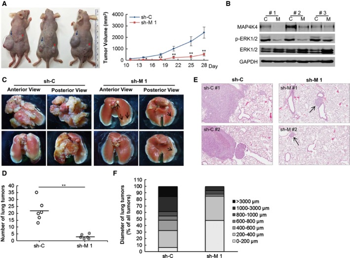
About 76% of patients with lung adenocarcinoma harbor activating mutations in the receptor tyrosine kinase (RTK)/RAS/RAF pathways, leading to aberrant activation of the mitogen-activated protein kinase (MAPK) pathways particularly the MAPK/ERK pathway. However, many lung adenocarcinomas lacking these genomic mutations also display significant MAPK pathway activation, suggesting that additional MAPK pathway alterations remain undetected. This study has identified serine/threonine kinase mitogen-activated protein 4 kinase 4 (MAP4K4) as a novel positive regulator of MAPK/ERK signaling in lung adenocarcinoma. The results showed that MAP4K4 was drastically elevated in lung adenocarcinoma independently of KRAS or EGFR mutation status. Knockdown of MAP4K4 inhibited proliferation, anchorage-independent growth and migration of lung adenocarcinoma cells, and also inhibited human lung adenocarcinoma xenograft growth and metastasis. Mechanistically, we found that MAP4K4 activated ERK through inhibiting protein phosphatase 2 activity. Our results further showed that downregulation of MAP4K4 prevented ERK reactivation in EGFR inhibitor erlotinib-treated lung adenocarcinoma cells. Together, our findings identify MAP4K4 as a novel MAPK/ERK pathway regulator in lung adenocarcinoma that is required for lung adenocarcinoma maintenance.

Keywords: EGFR; ERK; MAP4K4; cell signaling; lung adenocarcinoma.


Figures

Figure 1
Figure 1
KRAS or EGFR mutation‐independent elevation of MAP4K4 in lung adenocarcinomas. (A,B) Immunohistochemistry (IHC) staining against MAP4K4 was performed on TMA sections containing human lung adenocarcinoma (n = 44), adjacent normal lung tissue (n = 44), and normal lung tissue (n = 3) samples. Staining intensity was scored as 0, negative; 1, weak; 2, moderate; 3, strong. Representative pictures (A, left and middle panels) of distinct levels of MAP4K4 expression and a summary (B) of score distribution of all TMA samples were shown. Immunoblotting was performed to confirm the specificity of anti‐MAP4K4 antibody used in IHC staining (A, right panel). (C) MAP4K4 expression level was measured by IHC staining on TMA sections containing 136 lung adenocarcinoma samples with EGFR mutation, KRAS mutation, or wild‐type EGFR and KRAS. TMA slides were scored with the same method used in (A) and (B). Percentage of scored samples in three score groups was shown. P value was determined by chi‐square test; #denotes that there was not statistically significant difference (P > 0.05).
Figure 2
Figure 2
MAP4K4 is important for lung adenocarcinoma cell functions. MAP4K4‐knockdown cell lines or shRNA control cell lines were generated with lentiviral‐based shRNA targeting MAP4K4 (sh‐M 1 and sh‐M 2) or scrambled shRNA (sh‐C) in lung adenocarcinoma cell lines H23 (KRAS mutant), H1975 (EGFR mutant), and H1793 (KRAS and EGFR wild‐type). Data in line charts and column charts were shown as means ± SD, * and ** denote a statistically significant difference, P < 0.05 and P < 0.01, respectively, compared with parental cell lines. (A) Cell viability of different cell lines was measured by MTT assay. MAP4K4 expression was evaluated by immunoblotting (IB) at the time of plating. (B) Left panel: representative pictures of soft agar assay. Right panel: quantification of soft agar assay. (C) Left panel: representative pictures of wound healing assay using 24‐well wound healing inserts. Ten micromolar of mitomycin C was added to each well for 2 h after removing the inserts. Pictures were taken 0, 24, and 48 h after mitomycin C application. Right panel: quantification of wound healing assay. (D) Left panel: representative pictures of in vitro cell invasion assay. Right panel: quantification of in vitro cell invasion assay.
Figure 3
Figure 3
MAPK/ERK1/2 is a downstream signaling mediator of MAP4K4 in lung adenocarcinoma cells. (A) The whole‐cell lysates of different lung adenocarcinoma cell lines, including two KRAS‐mutant cell lines, A549 and H23; one KRAS and EGFR wild‐type cell line, H1793; three EGFR‐mutant cell lines, H1650, H1975, and H3255; and one lung bronchus cell line, BEAS‐2B, were used for IB with indicated antibodies. (B) MAP4K4‐knockdown cell lines (sh‐M 1 and sh‐M 2) or shRNA control cell lines (sh‐C) were generated with two different lentiviral‐based shRNA targeting MAP4K4 or scrambled shRNA in H23, H1975, and H1650 cell lines. The whole‐cell lysates were used for IB with indicated antibodies. To detect GTP‐bound RAS, the cell lysates were incubated with RAF‐1 RBD agarose. The bound proteins were then resolved by SDS/PAGE and blotted with anti‐RAS antibody. (C) MAP4K4‐overexpressing cell lines (HA‐M) and control cell lines (HA‐C) were established by transfecting pcDNA3.1‐HAMAP4K4 or pcDNA3.1‐HA into A549 or H3255 cell lines followed by G418 selection. The whole‐cell lysates were prepared for IB or subjected to RAS activation assay. (D–F) Constitutively active ERK2 (act ERK2) or vector was transfected into MAP4K4‐knockdown cell lines (sh‐M 1) with Polyjet In Vitro DNA Transfection Reagent. Data in column charts were shown as means ± SD; ** and # denote a statistically significant difference (P < 0.01) and no statistically significant difference (P > 0.05), respectively, compared with shRNA control cell lines (sh‐C). (D) Left panel: representative pictures of soft agar assay. Right panel: quantification of soft agar assay. (E) Left panel: representative pictures of in vitro cell invasion assay. Right panel: quantification of in vitro cell invasion assay. (F) The whole‐cell lysates of different cell lines were used for IB with indicated antibodies. (G) H1975‐sh‐control (sh‐C) and H1975‐sh‐MAP4K4 (sh‐M 1 and sh‐M 2) cells were treated with 3 μm of erlotinib for 6 and 24 h. IB was performed with indicated antibodies.
Figure 4
Figure 4
MAP4K4 regulates ERK activation through modulating PP2A activity. (A) The whole‐cell lysates of MAP4K4 shRNA‐knockdown or HA‐overexpressing, shRNA control or HA‐control, and their parental cell lines were used for IB with indicated antibodies. (B) PP2A activity of indicated cells was measured using PP2A immunoprecipitation phosphatase assay kit. * and ** denote a statistically significant difference, P < 0.05 and P < 0.01, respectively. (C) Whole‐cell lysates of H23 shRNA control cells (sh‐C), H23 MAP4K4‐knockdown cells (sh‐M 1) with and without PP2A inhibitor OA (5 nm) treatment for 24 h beforehand, A549 HA‐control cells (HA‐C) and A549 MAP4K4 HA‐overexpressing cells (HA‐M) with or without PP2A activator FTY720 (2.5 μm) treatment for 24 h beforehand were used for IB with indicated antibodies.
Figure 5
Figure 5
Downregulation of MAP4K4 inhibits in vivo xenograft growth and metastasis of lung adenocarcinoma cells. (A,B) 1 × 106 cells of H1975 shRNA control (sh‐C) and MAP4K4 shRNA‐knockdown cell lines (sh‐M 1) were injected subcutaneously into left and right flanks of the same nude mouse (n = 8). Mice were sacrificed 28 days after injection. (A) Left panel: picture of representative mice bearing lung adenocarcinoma xenograft. Dark blue arrows and red arrows indicated H1975‐sh‐control and H1975‐sh‐MAP4K4 xenografts, respectively. Right panel: Tumor volume change of each group was presented as means ± SD; * and ** denote a statistically significant difference, < 0.05 and P < 0.01, respectively, compared with shRNA control group. (B) IB of three pairs of representative xenograft tumors with indicated antibodies. Each pair was from the same mouse. C: H1975‐sh‐control xenograft; M: H1975‐sh‐MAP4K4 xenograft. (C–F) 1 × 106 cells of H1975‐sh‐control and H1975‐sh‐MAP4K4 cells were injected into tail vein of nude mice, six mice per group. Mice were sacrificed 12 weeks after injection. Lungs of each mouse were dissected and fixed with 10% formalin. (C) Pictures of representative lungs from each group were shown; black arrows indicate metastatic tumors. (D) Total number of surface lung tumors of all mice; ** denotes a statistically significant difference (P < 0.01) compared with shRNA control group. (E) Representative pictures of H&E‐stained slides of lung metastatic tumors of each group. (F) The largest diameter of each tumor observed on H&E‐stained slides was measured with ImageScope. The results were subdivided into different size groups. Percentage of the tumors within the group belonging to each subdivision was shown.

Similar articles

Cited by

References

    1. Ahearn IM, Haigis K, Bar‐Sagi D and Philips MR (2012) Regulating the regulator: post‐translational modification of RAS. Nat Rev Mol Cell Biol 13, 39–51. - PMC - PubMed
    1. Aouadi M, Tesz GJ, Nicoloro SM, Wang M, Chouinard M, Soto E, Ostroff GR and Czech MP (2009) Orally delivered siRNA targeting macrophage Map4k4 suppresses systemic inflammation. Nature 458, 1180–1184. - PMC - PubMed
    1. Bae D and Ceryak S (2009) Raf‐independent, PP2A‐dependent MEK activation in response to ERK silencing. Biochem Biophys Res Commun 385, 523–527. - PMC - PubMed
    1. Bossi D, Cicalese A, Dellino GI, Luzi L, Riva L, D'Alesio C, Diaferia GR, Carugo A, Cavallaro E, Piccioni R et al (2016) In vivo genetic screens of patient‐derived tumors revealed unexpected frailty of the transformed phenotype. Cancer Discov 6, 650–663. - PubMed
    1. Bouzakri K, Ribaux P and Halban PA (2009) Silencing mitogen‐activated protein 4 kinase 4 (MAP4K4) protects beta cells from tumor necrosis factor‐alpha‐induced decrease of IRS‐2 and inhibition of glucose‐stimulated insulin secretion. J Biol Chem 284, 27892–27898. - PMC - PubMed

MeSH terms

Substances