Review
. 2017 May;26(3):154-164.
doi: 10.1097/MNH.0000000000000311.

Can podocytes be regenerated in adults?

Affiliations
Review

Can podocytes be regenerated in adults?

Stuart J Shankland et al. Curr Opin Nephrol Hypertens. 2017 May.

Abstract

Purpose of review: Podocytes are critical components of the nephron filtration barrier and are depleted in many kidney injuries and disease states. Terminally differentiated adult podocytes are highly specialized, postmitotic cells, raising the question of whether the body has any ability to regenerate lost podocytes. This timely question has recently been illuminated by a series of innovative studies. Here, we review recent progress on this topic of significant interest and debate.

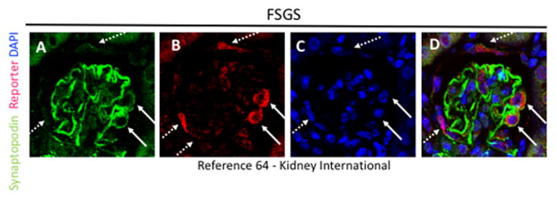
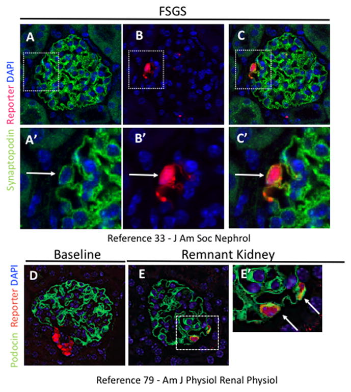
Recent findings: The innovation of genetic labeling techniques enables fate tracing of individual podocytes, providing the strongest evidence yet that podocytes can be replaced by nearby progenitor cells. In particular, two progenitor pools have recently been identified in multiple studies: parietal epithelial cells and cells of renin lineage. These studies furthermore suggest that podocyte regeneration can be enhanced using ex-vivo or pharmacological interventions.

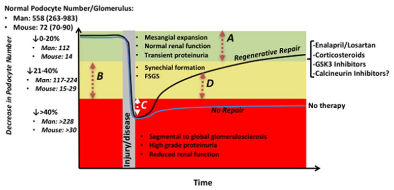
Summary: Recent studies indicate that the podocyte compartment is more dynamic than previously believed. Bidirectional exchange with neighboring cellular compartments provides a mechanism for podocyte replacement. Based on these findings, we propose a set of criteria for evaluating podocyte regeneration and suggest that restoration of podocyte number to a subsclerotic threshold be targeted as a potentially achievable clinical goal.


Conflict of interest statement

COI: None of the authors have any financial or other conflicts of interest.

Figures

Figure 1
Figure 1. Reporter labeled PECs migrate onto the glomerular tuft in FSGS, and express synaptopodin
(A) Synaptopodin staining (green) is restricted to podocytes. (B) β-gal staining (red) is detected in cells lining Bowman’s capsule (dashed arrows) and inside the tuft (solid arrows). (C) DAPI staining is restricted to nuclei (blue). (D) Merge of all three stains, showing that a subset of reporter labeled PECs on the tuft co-stain with synaptopodin (solid arrows).
Figure 2
Figure 2. Reporter labeled CoRL migrate onto the glomerular tuft in FSGS and remnant kidney, and express synaptopodin and podocin
(A) Synaptopodin staining (green) is restricted to podocytes. (B) RFP staining (red) is detected within glomerular tuft (white arrow). (C) Synaptopodin and RFP staining colocalize (white arrow) in the glomerular tuft. (A′–C′) Higher magnification of insets shown above. (D) At baseline, podocin staining (green) is restricted to podocytes and RFP staining (red) is restricted to CoRL in the juxtaglomerulus. (E) In the remnant kidney model, reporter labeled CoRL on the glomerular tuft co-stain with podocin. (E′) Higher magnification of inset shown in E (white arrows indicate co-staining).
Figure 3
Figure 3. Podocyte loss and regeneration, and their relationship to structural glomerular changes
The Y axis shows the normal podocyte number and range per glomerulus in man and mouse. Three scenarios represented by different colors are shown for podocyte depletion, based on studies by Wharram.8 A decrease in podocyte number by 20% (represented by the green area) corresponds to a decrease in podocyte number per glomerulus by 112 in man, and 14 in mice. A decrease in podocyte number by 21–40% (represented by the yellow area) corresponds to a decrease in podocyte number per glomerulus by 117–224 in man, and 15–29 in mice. A decrease in podocyte number by greater than 40% (represented by the red area) corresponds to a decrease in podocyte number per glomerulus by >228 in man, and >30 in mice. A decrease in podocyte number by 20% (represented by the red dashed arrow A), is not accompanied by any serious decline in kidney function, at least over the short term, unless there is subsequent podocyte depletion. In a steady state, we argue therefore that there is no major biological need to replace these podocytes. There is however a need to replace podocytes when their number is depleted between 21–40%, in order to limit further synchial formation and focal segmental glomerulosclerosis (FSGS), and to even reverse these processes. However, podocyte replacement does NOT need to return to the normal number in order for this to happen. Rather, we propose that the goal to replace podocyte number is above the threshold for scarring. As shown in the graph, this is typically less than 20% of normal (represented by the dashed arrow B). For example, to reach 20% of normal from a nadir of 40% of normal to prevent/reverse scarring in an individual glomerulus, requires only a 20% increase in podocyte number for that glomerulus (red arrow B). The mean number of glomeruli in a mouse kidney is 12,083±1009 per kidney, or 24,166 glomeruli total per mouse.80 In FSGS for example, if 20% of glomeruli are affected, the number of podocytes that need to be replaced in one kidney to return to the original baseline is 67,665 (20% of glomeruli * 24,166 glomeruli * 14 podocytes lost per glomerulus). If podocyte number drops by 40% (decrease of 29 podocytes in a single glomerulus), global glomerulosclerosis will ensue. Thus, still using the example of 20% of glomeruli affected implies that 20 % of glomeruli *24,166 glomeruli *29 podocytes lost per glomerulus= 140,162 total podocytes needed. Thus, for repair from a 40% drop in podocytes, to a 20% drop in podocytes (above which scarring does not occur), 72,497 podocytes are needed. While complete restoration would require 140,162 podocytes. If podocyte number drops below the 40% threshold to 50% for example, then an immediate goal for replacement would be to increase podocyte number by 10% (white arrow C) in order to limit the progression from focal to global sclerosis, and to reverse global to focal sclerosis. The second goal is to increase podocyte number by another 20% to limit focal sclerosis (red arrow B). Finally, several therapies (shown on the right side of the graph) increase podocyte number and in doing so, limit and/or reverse the progression of scarring by regenerative repair (black line)(example is red arrow D), whereas no therapy leads to non-regenerative repair (blue line)

Similar articles

Cited by

References

    1. Little MH. The Life Cycle of the Nephron Progenitor. Dev Cell. 2015;35(1):5–6. - PubMed
    1. Little MH, McMahon AP. Mammalian kidney development: principles, progress, and projections. Cold Spring Harb Perspect Biol. 2012;4(5) - PMC - PubMed
    1. Hartman HA, Lai HL, Patterson LT. Cessation of renal morphogenesis in mice. Dev Biol. 2007;310(2):379–387. - PMC - PubMed
    1. Kobayashi A, Valerius MT, Mugford JW, Carroll TJ, Self M, Oliver G, McMahon AP. Six2 defines and regulates a multipotent self-renewing nephron progenitor population throughout mammalian kidney development. Cell Stem Cell. 2008;3(2):169–181. - PMC - PubMed
    1. Ashraf S, Gee HY, Woerner S, Xie LX, Vega-Warner V, Lovric S, Fang H, Song X, Cattran DC, Avila-Casado C, Paterson AD, Nitschke P, Bole-Feysot C, et al. ADCK4 mutations promote steroid-resistant nephrotic syndrome through CoQ10 biosynthesis disruption. J Clin Invest. 2013;123(12):5179–5189. - PMC - PubMed