Review
. 2017 Jun;38(5):774-779.
doi: 10.1097/MAO.0000000000001377.

Secondary Endolymphatic Hydrops

Affiliations
Review

Secondary Endolymphatic Hydrops

Ashley P O'Connell Ferster et al. Otol Neurotol. 2017 Jun.

Abstract

Hypothesis: A review of the most recent literature will provide clinicians with an update of secondary endolymphatic hydrops, aiding in diagnosis and treatment of affected patients.

Background: Secondary endolymphatic hydrops is a pathologic finding of the inner ear resulting in episodic vertigo and intermittent hearing loss. It is a finding for which extensive research is being performed.

Methods: A review of the most recent literature on secondary endolymphatic hydrops was performed using PubMed literature search.

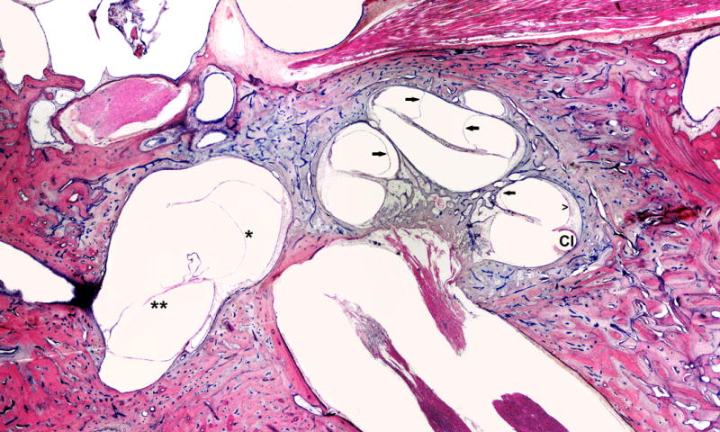
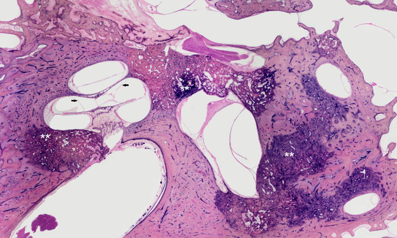
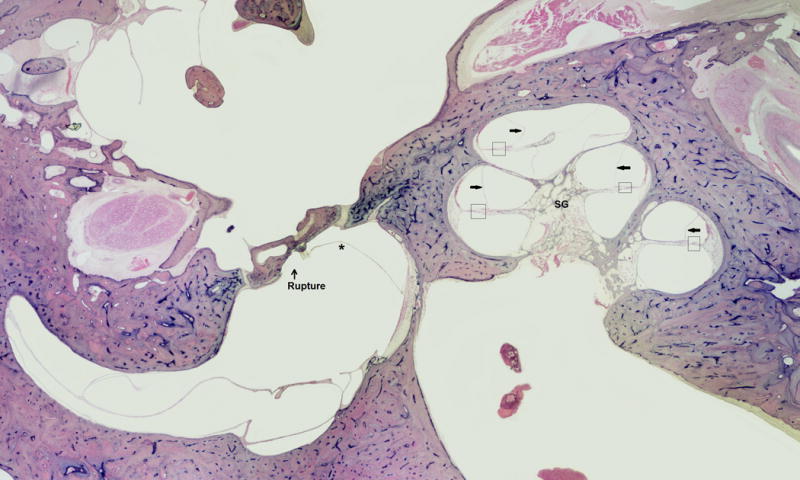
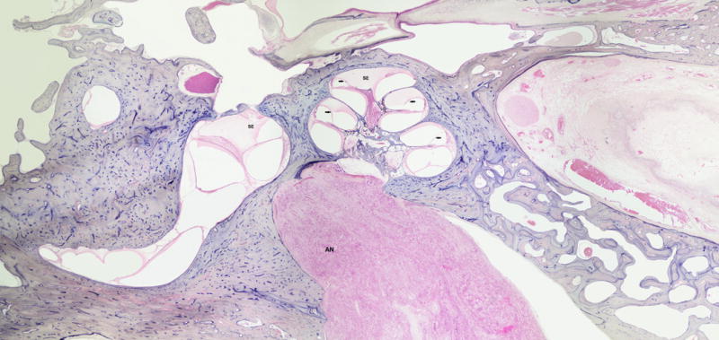
Results: Recent investigation of secondary endolymphatic hydrops has brought attention to traumatic and inflammatory insults as causes for secondary endolymphatic hydrops. Such etiologies, including postsurgical effects of cochlear implantation and endolymphatic sac ablation; otosclerosis and its operative intervention(s); acoustic and mechanical trauma; medications; and systemic inflammatory processes, have been determined as causes of secondary lymphatic hydrops. Histopathological slides for many of the etiologies of secondary endolymphatic hydrops are presented.

Conclusion: Through an understanding of the pathophysiology and etiologies of secondary endolymphatic hydrops, clinicians will gain a better understanding of this complex disease process, which will aid in treatment of patients with this disease process.


Conflict of interest statement

Conflicts of Interest: None

Figures

FIG. 1
FIG. 1
This is a 66-year-old white male with a clinical history of heart attack, occupational noise exposure, and left sensorineural hearing loss. He was treated with left cochlear implant (CI). Histopathology evaluation of the left ear showed: Simple mastoidectomy cavity, profound hydrops (arrows) in all turns of cochlea, saccular (*) and utricular hydrops (**), atrophy of stria vascularis (open arrow head) in lower basal turn, and hyalinization and fibrosis around electrode insertion site (CI).
FIG. 2
FIG. 2
This is a 78-year-old white female with a clinical history of bilateral profound hearing loss, as well as left tympanotomy and stapedectomy. She became extremely vertiginous in the post-operative period. Histopathologic examination showed: LEFT: stapedectomy, otosclerosis, dehiscent carotid artery and facial nerve, and profound saccular hydrops (*), decreased ganglion cells (SG).
FIG. 3
FIG. 3
This is a 78-year-old female patient with a history of Meniere’s Disease and decreased hearing. Histopathologic examination showed otosclerosis, cochlear (arrows) and utricular hydrops. Note that otosclerosis blocks the endolymphatic duct.
FIG. 4
FIG. 4
This is an 86-year-old male with a clinical history of Meniere’s disease and a Tack operation. Histopathology of the ear showed Tack hole seen in stapes footplate, severe endolymphatic hydrops, saccule hydropic and ruptured, severe loss ganglion cells, strial atrophy, and organ of Corti atrophy.
FIG. 5
FIG. 5
This is a 76-year-old black male with a clinical history of dizziness and severe sensorineural hearing loss. Histopathologic evaluation of the left ear showed intracanalicular vestibular schwannoma (AN), serous labyrinthitis (SE), and cochlear hydrops (arrows).
FIG. 6
FIG. 6
This is a 29-year-old white male with a clinical history of Leukemia, dizziness, and left severe sensorineural hearing loss. His hearing loss was fluctuant. Histopathologic evaluation of the ear showed: hypertrophied middle ear mucosa, fibrous tissues in the posterior side of the middle ear, profound endolymphatic hydrops (arrows), severe loss of haircells, and strial atrophy (open arrow head).
FIG. 7
FIG. 7
This is a 16-year-old white, male with leukemia, dizziness and right hemotympanum. Histopathologic examination of the ear showed leukemic hemorrhage in the middle and inner ears with cochlear (arrows) and slight saccular hydrops (*).

References

    1. Salt AN, Plontke SK. Endolymphatic hydrops: pathophysiology and experimental models. Otolaryngol Clin North Am. 2010;43:971–83. - PMC - PubMed
    1. Chen YJ, Young YH. Secondary endolymphatic hydrops after acoustic trauma. Otol Neurol. 2016;36:1–6. - PubMed
    1. Fontaine N, Chapiot A, Debry C, Gentine A. A case of spontaneous intracranial hypotension: from Meniere-like syndrome to cerebral involvement. Euro Ann Otorhinolaryngol Head Neck Dis. 2012;129:153–6. - PubMed
    1. Smeds H, Eastwood HT, Hapson AJ, Sale P, Campbell LJ, Arhatari BD, Mansour S, O’Leary SJ. Endolymphatic hydrops is prevalent in the first weeks following cochlear implantation. Hearing Res. 2015;327:48–57. - PubMed
    1. Handzel O, Burgess BJ, Nadol JB. Histopathology of the peripheral vestibular system after cochlear implantation in the human. Otol Neurotol. 2006;27:57 e64. - PubMed