. 2017 Mar 17;12(3):e0173715.
doi: 10.1371/journal.pone.0173715. eCollection 2017.

Hypervirulent Mycobacterium tuberculosis strain triggers necrotic lung pathology associated with enhanced recruitment of neutrophils in resistant C57BL/6 mice

Affiliations

Hypervirulent Mycobacterium tuberculosis strain triggers necrotic lung pathology associated with enhanced recruitment of neutrophils in resistant C57BL/6 mice

Fabrício M Almeida et al. PLoS One. .

Erratum in

Abstract

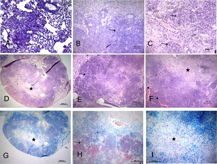
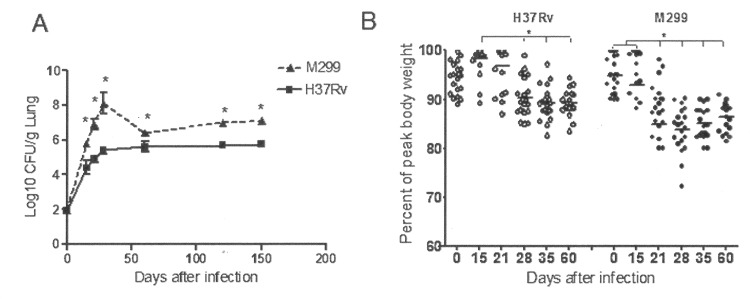
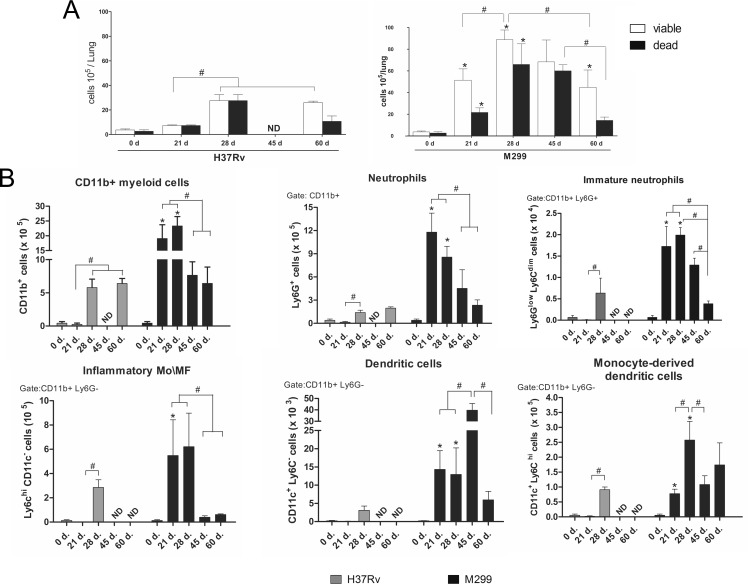
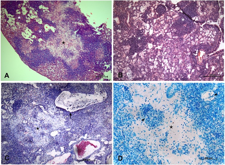
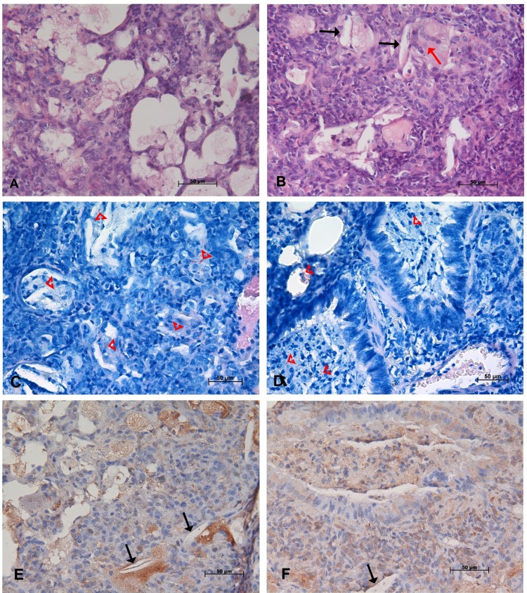
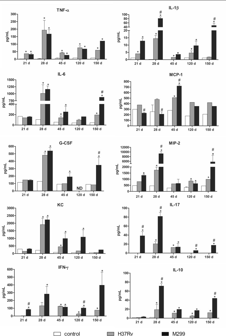
Tuberculosis (TB) is a chronic infectious disease caused by Mycobacterium tuberculosis (Mtb) that in most cases induces irreversible necrosis of lung tissue as a result of excessive inflammatory reactions. The murine model of TB in resistant C57BL/6 mice infected with reference Mtb strains is widely used in TB studies; however, these mice do not show a necrotic pathology, which restricts their use in studies of irreversible tissue damage. Recently, we demonstrated that necrotic lung lesions could be induced in the C57BL/6 mice by highly virulent Mtb strains belonging to the modern Beijing sublineage. However, the pathogenic mechanisms leading to necrosis in this model were not elucidated. In this study, we investigated the dynamics of lung lesions in mice infected with highly virulent Beijing Mtb strain M299, compared with those infected with laboratory Mtb strain H37Rv. The data demonstrate that necrotic lung lesions in mice infected by the strain M299 were associated with enhanced recruitment of myeloid cells, especially neutrophils, and increased levels of proinflammatory cytokines, consistent with exacerbated inflammation. High levels of IFN-γ production contributed to the control of bacterial growth. Further progression to chronic disease was associated with a reduction in the levels of inflammatory mediators in the lungs, the accumulation of foamy macrophages and partial healing of the necrotic tissue by fibrosis. At a late stage of disease, degradation of foamy cells resulted in the liberation of accumulated lipids and persisting bacilli and further activation of inflammation, which promoted lung consolidation. Overall, our studies show that C57BL/6 mice infected with highly virulent Mtb strain may serve as a TB model reproducing an exacerbated inflammatory response in a resistant host to hypervirulent mycobacteria, leading to irreversible necrotic lung lesions.


Conflict of interest statement

Competing Interests: The authors have declared that no competing interests exist.

Figures

Fig 1
Fig 1. Kinetics of lung histopathology during the acute stage of infection in C57BL/6 mice inoculated with strain M299.
Mice were intratracheally infected with ~100 CFU. Pathologic alterations of lung tissue were determined on day 15 p.i. (A), day 21 p.i. (B, C) and day 28 p.i. (D- I) by microscopy of lung sections stained with hematoxylin-eosin (A- F) and Ziehl-Neelsen (G- I). Panel A shows interstitial granulomatous infiltrates on day 15 p.i. Panels B and C show the development of alveolitis through infiltration of alveoli (black arrows) with inflammatory cells, predominantly by neutrophils (enclosed in black circles). Panels D and G show coalescing foci of tuberculous pneumonia leading to the formation of tubercules with extensive areas of central caseous necrosis (black stars). The peripheral zone of the necrotic lesion is amplified in E and H, demonstrating areas of alveolitis (black arrows) with thickened alveolar walls and intra-alveolar cellular exudates composed predominantly of neutrophil debris (E) and numerous AFB (H). The caseous core is amplified in F and I. Note the thrombosed septal capillaries (black arrow heads) and obstruction of small airways with cellular debris (white arrow) in the peripheral rim of the necrotic zone (F). Numerous extracellular AFB and clumps of bacteria can be seen in the necrotic area (I). Data are representative of four independent experiments. Bars represent 500 μm—in the panels D and G, 200 μm—in B, E and H, 100 μm—in A, F and I and 20 μm—in C.
Fig 2
Fig 2. Kinetics of bacterial growth in the lungs and morbidity of C57BL/6 mice inoculated with Mtb strains.
Mice were infected i.t. with 100 CFU of the hypervirulent strain M299 and laboratory strain H37Rv. A. Bacterial burdens in the lungs were quantified by CFU counting on days 0, 5, 21, 28, 120 and 150 after infection. Data from three experiments (n = 3 for each group and each experiment) are expressed logarithmically as the mean log10 CFU standard deviation (SD) (error bars). Mean values that were significantly different from the respective mean value of the group infected by strain H37Rv are indicated by asterisks *, p < 0.05. B. Kinetics of post-infection changes in mouse body weight. Weight loss was used as an indicator of morbidity. Data are presented as the percentage of peak body weight of each mice (n = 20, pooled data from two independent experiments). Statistical differences are indicated by asterisks *, p < 0.05.
Fig 3
Fig 3. Kinetics of myeloid cell recruitment into the lungs of mice infected with strains M299 and H37Rv.
A. Total numbers of cells recovered from the lungs over a follow-up period of 60 days after inoculation with bacilli or sterile PBS (control, day 0). Proportions of viable and dead cells were determined by trypan-blue staining. B. Myeloid cell populations were identified by flow cytometry. Total numbers of CD11b+ myeloid cells per lung and individual myeloid cell subpopulations, including neutrophils (CD11b+Ly6G+Ly6C+cells), immature neutrophil precursors (CD11b+Ly6GlowLy6Cdim), inflammatory monocytes and macrophages (CD11b+Ly6ChiCD11c-Ly6G- cells), monocyte-derived dendritic cells (CD11b+CD11c+Ly6ChiLy6G-) and dendritic cells (CD11b+CD11c+Ly6C-Ly6G-), were calculated. Values shown are the mean numbers of cells per lung ± SD, n = 8–9 lungs at each time point, pooled data from three independent experiments. Mean values of groups infected by strain M299 that were significantly different from the respective mean value of groups infected by H37Rv strain are indicated by asterisks * (p < 0.05). Significant differences between groups infected by each strain are indicated by lines and octothorpes #, (p < 0.05).
Fig 4
Fig 4. Lung histopathology at the early chronic stage of infection by highly virulent Mtb strain M299.
Pathological alterations to lung tissue were observed on day 120 after infection by microscopic analysis of lung sections stained by hematoxylin-eosin (A and B), Masson´s trichrome (C) and Ziehl-Neelsen (D). Panel A demonstrates a necrotic lesion (black star) partially healed by fibrosis and surrounded by a rim of lymphocytes and lipid-laden foamy macrophages in the external region. Secondary granulomatous lesions are marked by white stars. Panel B demonstrates a higher-magnification image of two compact perivascular lymphocyte granulomas, surrounded by foamy macrophages. C. Masson’s staining revealed the presence of profuse collagen (in light-blue) in the central area of the necrotic lesion. Note cellular debris in the bronchiole and the absence of a bronchiolar epithelium (black arrow). D. Single AFB, exhibiting weak staining (black arrow heads), could be seen in necrotic areas partially healed by fibrosis and in foamy macrophages within alveoli. Bars represent 500 μm- in the panel A, 200 μm- in the panels B and C; and 100 μm- in the panel D.
Fig 5
Fig 5. Lung histopathology at the late chronic stage of infection by highly virulent strain M299.
Pathological alterations to lung tissue were observed on day 150 after infection by microscopic analysis of lung sections stained by hematoxylin-eosin (A and B), Ziehl-Neelsen (C and D) and immunohistochemical staining method for mycobacterial antigens (E and F). Panels A and B demonstrate massive intra-alveolar disintegration of foamy macrophages admixed with recruited neutrophils and appearance of multinucleated cells (red arrow, B). The resulting cell debris filled small airways (D and F). Numerous AFB were seen in foamy macrophages and cell debris (red arrowheads, C and D). Necrotic death of foamy cells led to the liberation of mycobacterial antigens (stained in brown) and the accumulation of needle-shaped cholesterol clefts (black arrows, B, E and F). Bars represent 50 μm.
Fig 6
Fig 6. Production of cytokines and chemokines by cells obtained from the lungs of C57BL/6 mice infected with strains M299 and H37Rv.
Lung cells were isolated on days 21, 28, 45, 120 and 150 after i.t. infection and cultured in 96-well plates, 5 x 10⁴ cells/well. Culture supernatants were collected after 48 h of incubation and cytokine concentrations were measured by the Multiplex method. The data were obtained in three independent experiments. Significant differences between infected and uninfected groups are indicated by asterisks * (p <0.05). Mean values of groups infected by strain M299 that were significantly different from the respective mean value of groups infected by strain H37Rv are indicated by octothorpes # (p < 0.05). ND- not defined.

References

    1. Orme IM. A new unifying theory of the pathogenesis of tuberculosis. Tuberculosis (Edinb). 2014;94(1):8–14. - PMC - PubMed
    1. Orme IM. The mouse as a useful model of tuberculosis. Tuberculosis (Edinb). 2003; 83(1–3):112–5. - PubMed
    1. Kramnik I, Dietrich WF, Demant P, Bloom BR: Genetic control of resistance to experimental infection with virulent Mycobacterium tuberculosis. Proc Natl Acad Sci USA. 2000;97:8560–65. 10.1073/pnas.150227197 - DOI - PMC - PubMed
    1. Di Pietrantonio T, Schurr E. Mouse models for the genetic study of tuberculosis susceptibility. Briefings in functional genomics and proteomics. 2005;4(3):277–92. - PubMed
    1. Keller C, Hoffmann R, Lang R, Brandau S, Hermann C, Ehlers S. Genetically determined susceptibility to tuberculosis in mice causally involves accelerated and enhanced recruitment of granulocytes. Infect Immun. 2006;74(7):4295–309. 10.1128/IAI.00057-06 - DOI - PMC - PubMed

MeSH terms