. 2017 Oct;33(10):1018-1026.
doi: 10.1089/AID.2016.0030. Epub 2017 Apr 24.

Dysregulation of Alveolar Macrophage PPARγ, NADPH Oxidases, and TGFβ1 in Otherwise Healthy HIV-Infected Individuals

Affiliations

Dysregulation of Alveolar Macrophage PPARγ, NADPH Oxidases, and TGFβ1 in Otherwise Healthy HIV-Infected Individuals

Samantha M Yeligar et al. AIDS Res Hum Retroviruses. 2017 Oct.

Abstract

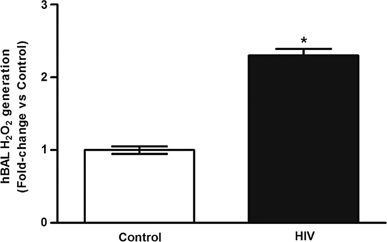
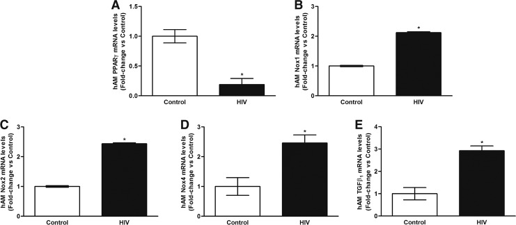
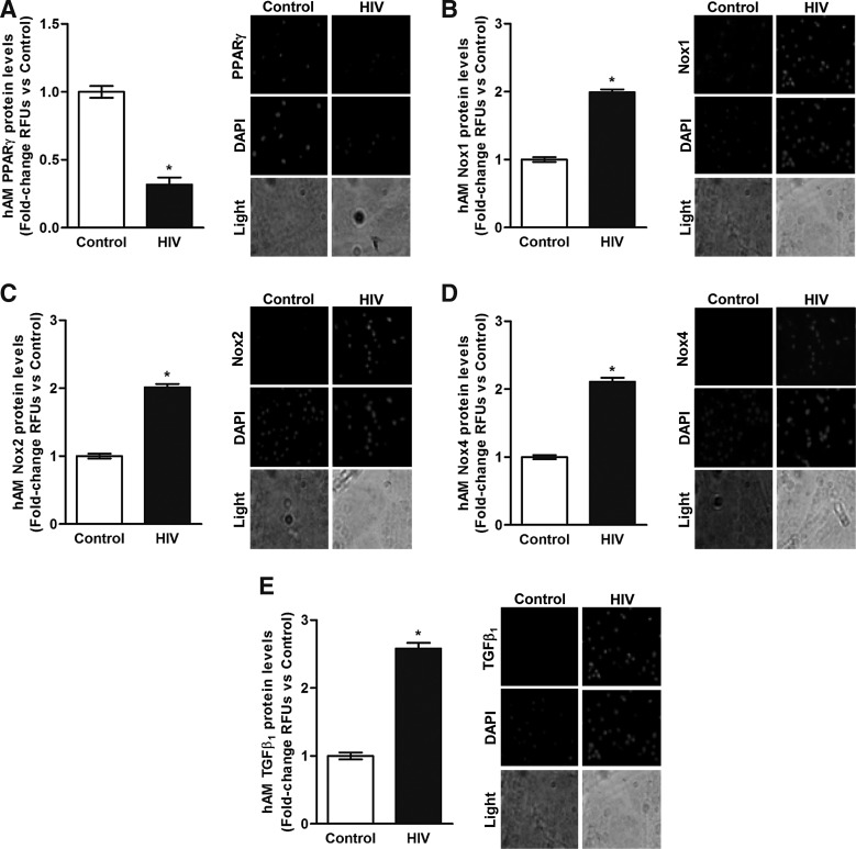
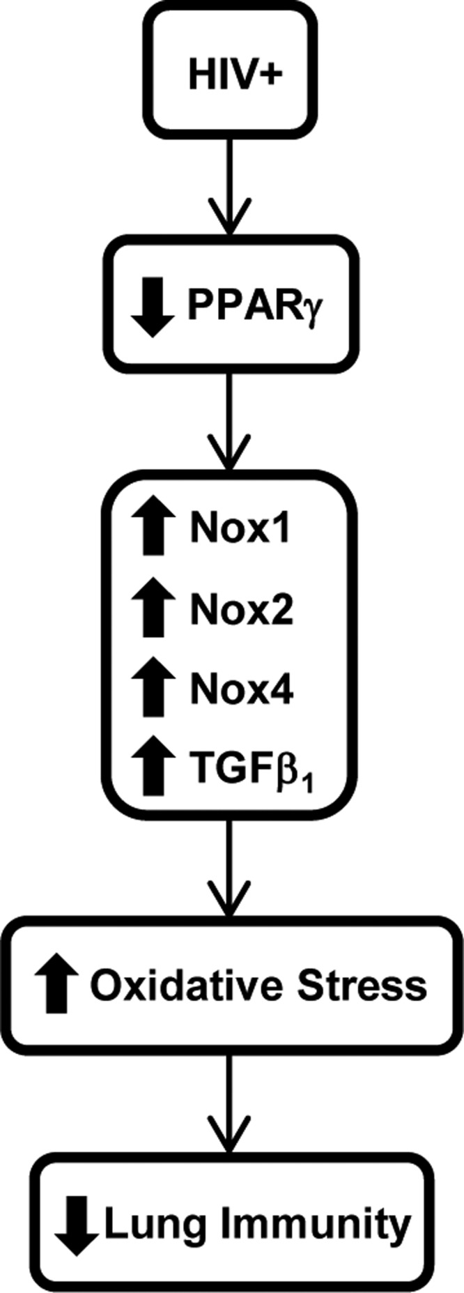
Despite antiretroviral therapy (ART), respiratory infections increase mortality in individuals living with chronic human immunodeficiency virus (HIV) infection. In experimental and clinical studies of chronic HIV infection, alveolar macrophages (AMs) exhibit impaired phagocytosis and bacterial clearance. Peroxisome proliferator-activated receptor (PPAR)γ, NADPH oxidase (Nox) isoforms Nox1, Nox2, Nox4, and transforming growth factor-beta 1 (TGFβ1) are critical mediators of AM oxidative stress and phagocytic dysfunction. Therefore, we hypothesized that HIV alters AM expression of these targets, resulting in chronic lung oxidative stress and subsequent immune dysfunction. A cross-sectional study of HIV-infected (n = 22) and HIV-uninfected (n = 6) subjects was conducted. Bronchoalveolar lavage (BAL) was performed, and AMs were isolated. Lung H2O2 generation was determined by measuring H2O2 in the BAL fluid. In AMs, PPARγ, Nox1, Nox2, Nox4, and TGFβ1 mRNA (quantitative real-time polymerase chain reaction) and protein (fluorescent immunomicroscopy) levels were assessed. Compared with HIV-uninfected (control) subjects, HIV-infected subjects were relatively older and the majority were African American; ∼86% were on ART, and their median CD4 count was 445, with a median viral load of 0 log copies/ml. HIV infection was associated with increased H2O2 in the BAL, decreased AM mRNA and protein levels of PPARγ, and increased AM mRNA and protein levels of Nox1, Nox2, Nox4, and TGFβ1. PPARγ attenuation and increases in Nox1, Nox2, Nox4, and TGFβ1 contribute to AM oxidative stress and immune dysfunction in the AMs of otherwise healthy HIV-infected subjects. These findings provide novel insights into the molecular mechanisms by which HIV increases susceptibility to pulmonary infections.

Keywords: HIV; immunology; molecular biology.


Conflict of interest statement

No competing financial interests exist.

Figures

<b>FIG. 1.</b>
FIG. 1.
Chronic HIV infection is associated with increases in BAL oxidative stress. BAL fluid was collected from HIV-uninfected control subjects (Control, n = 6) and from HIV-infected subjects (HIV, n = 22). H2O2 generation was measured by Amplex Red assay (n = 28, in duplicate). Values were normalized to total protein and are expressed as mean ± SEM, relative to control. *p < .05 versus control. BAL, bronchoalveolar lavage; SEM, standard error of the mean.
<b>FIG. 2.</b>
FIG. 2.
Chronic HIV infection is associated with alterations in mRNA levels of PPARγ, Nox1, Nox2, Nox4, and TGFβ1 in AMs. AMs were collected from HIV-uninfected control subjects (Control, n = 6) and from HIV-infected subjects (HIV, n = 22). mRNA levels of PPARγ (A), Nox1 (B), Nox2 (C), Nox4 (D), and TGFβ1 (E) were measured by quantitative real-time polymerase chain reaction (n = 28, in duplicate), normalized to 9s mRNA, and expressed as mean ± SEM, relative to control. *p < .05 versus control. AMs, alveolar macrophages; PPARγ, peroxisome proliferator-activated receptor gamma; TGFβ1, transforming growth factor beta-1.
<b>FIG. 3.</b>
FIG. 3.
Chronic HIV infection is associated with alterations in protein levels of PPARγ, Nox1, Nox2, Nox4, and TGFβ1 in AMs. AMs were collected from HIV-uninfected control subjects (Control, n = 6) and from HIV-infected subjects (HIV, n = 22). Protein levels of PPARγ (A), Nox1 (B), Nox2 (C), Nox4 (D), and TGFβ1 (E) were determined in AMs (n = 28, 10 fields per experimental condition) by using confocal fluorescence microscopy. Representative images are provided. Quantification of fluorescence values was normalized to DAPI nuclear stain and expressed as mean relative fluorescence units (RFU) per cell ± SEM, relative to control. *p < .05 versus control.
<b>FIG. 4.</b>
FIG. 4.
Ex vivo PIO treatment reverses chronic HIV infection-associated alterations in mRNA and protein levels of PPARγ, Nox1, Nox2, Nox4, and TGFβ1 and improves phagocytosis in AMs. AMs were collected from HIV-infected subjects and were treated with PIO (HIV + PIO) or dimethyl sulfoxide vehicle (HIV) (n = 4). mRNA levels of PPARγ (A), Nox1 (B), Nox2 (C), Nox4 (D), and TGFβ1 (E) were measured by qRT-PCR (n = 8, in duplicate), normalized to 9s mRNA, and expressed as mean ± SEM, relative to HIV. AM phagocytic ability (F) was assessed by phagocytosis assay and fluorescent microscopy imaging (10 fields per experimental condition). Phagocytic index was calculated as the percentage of phagocytic cells multiplied by the relative fluorescence units of S. aureus per cell. All values were expressed as mean ± SEM, relative to HIV. *p < .05 versus HIV. PIO, pioglitazone.
<b>FIG. 5.</b>
FIG. 5.
Hypothetical schema of the molecular mechanisms involved in HIV-associated lung immune dysfunction. Chronic HIV infection is associated with impaired lung immunity through decreases in AMs PPARγ expression. Attenuation of PPARγ increases AMs expression of Nox isoforms, and TGFβ1, which, in turn, enhances lung oxidative stress. This increase in lung oxidative stress causes the lung immune dysfunction observed in HIV-infected individuals.

References

    1. Li TS, Tubiana R, Katlama C, Calvez V, Ait Mohand H, Autran B: Long-lasting recovery in CD4 T-cell function and viral-load reduction after highly active antiretroviral therapy in advanced HIV-1 disease. Lancet 1998;351:1682–1686 - PubMed
    1. Mellors JW, Munoz A, Giorgi JV, Margolick JB, Tassoni CJ, Gupta P, et al. : Plasma viral load and CD4+ lymphocytes as prognostic markers of HIV-1 infection. Ann Intern Med 1997;126:946–954 - PubMed
    1. Gordin FM, Roediger MP, Girard PM, Lundgren JD, Miro JM, Palfreeman A, et al. : Pneumonia in HIV-infected persons: Increased risk with cigarette smoking and treatment interruption. Am J Respir Crit Care Med 2008;178:630–636 - PMC - PubMed
    1. Jordano Q, Falco V, Almirante B, Planes AM, del Valle O, Ribera E, et al. : Invasive pneumococcal disease in patients infected with HIV: Still a threat in the era of highly active antiretroviral therapy. Clin Infect Dis 2004;38:1623–1628 - PubMed
    1. Strategies for Management of Antiretroviral Therapy Study Group; El-Sadr WM, Lundgren J, Neaton JD, Gordin F, Abrams D, et al. : CD4+ count-guided interruption of antiretroviral treatment. N Engl J Med 2006;355:2283–2296 - PubMed

MeSH terms