Review
. 2017 May 1;312(5):H1052-H1059.
doi: 10.1152/ajpheart.00024.2017. Epub 2017 Mar 17.

Immune checkpoint dysfunction in large and medium vessel vasculitis

Affiliations
Review

Immune checkpoint dysfunction in large and medium vessel vasculitis

Ryu Watanabe et al. Am J Physiol Heart Circ Physiol. .

Abstract

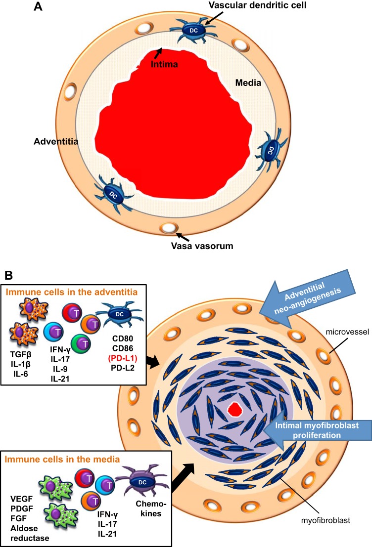
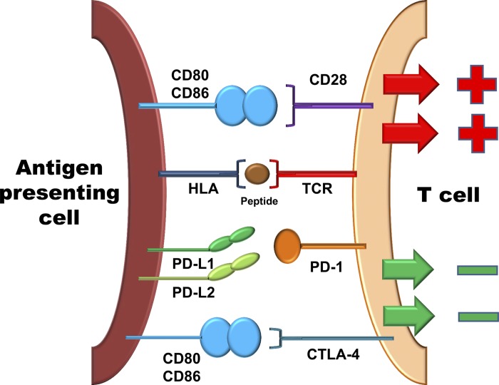
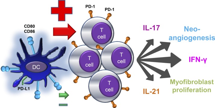
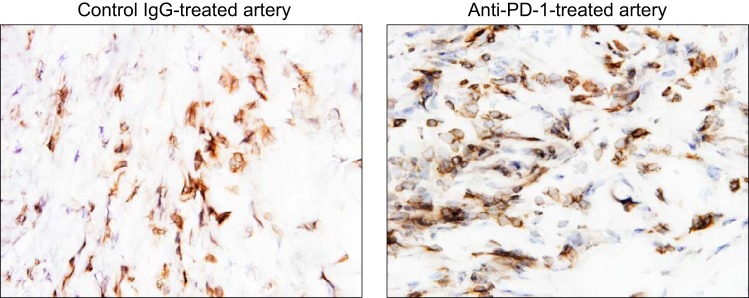
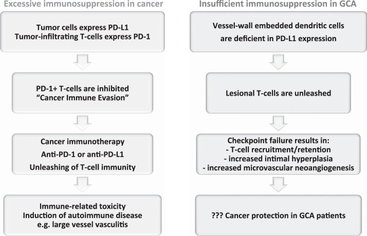
Giant cell arteritis (GCA) is a granulomatous vasculitis of the aorta and its medium-sized branch vessels. CD4 T cells, macrophages, and dendritic cells (DCs) build granulomatous infiltrates that injure the vessel wall and elicit a maladaptive response to injury. Pathological consequences include fragmentation of elastic membranes, destruction of the medial layer, microvascular neoangiogenesis, massive outgrowth of myofibroblasts, and lumen-occlusive intimal hyperplasia. Antigens have been suspected to drive the local activation of vasculitogenic CD4 T cells, but recent data have suggested a more generalized defect in the threshold setting of such T cells, rendering them hyperreactive. Under physiological conditions, immune checkpoints provide negative signals to curb T cell activation and prevent inflammation-associated tissue destruction. This protective mechanism is disrupted in GCA. Vessel wall DCs fail to express the immunoinhibitory ligand programmed cell death ligand-1, leaving lesional T cells unchecked. Consequently, programmed cell death protein-1-positive CD4 T cells can enter the immunoprivileged vessel wall, where they produce a broad spectrum of inflammatory cytokines (interferon-γ, IL-17, and IL-21) and have a direct role in driving intimal hyperplasia and intramural neoangiogenesis. The deficiency of the programmed cell death protein-1 immune checkpoint in GCA, promoting unopposed T cell immunity, contrasts with checkpoint hyperactivity in cancer patients in whom excessive programmed cell death ligand-1 expression paralyzes the function of antitumor T cells. Excessive checkpoint activity is the principle underlying cancer-immune evasion and is therapeutically targeted by immunotherapy with checkpoint inhibitors. Such checkpoint inhibitors, which unleash anticancer T cells and induce immune-related toxicity, may lead to drug-induced vasculitis.

Keywords: dendritic cell; giant cell arteritis; immune checkpoint; programmed cell death ligand-1; programmed cell death protein-1.


Figures

Fig. 1.
Fig. 1.
Immune lesion in giant cell arteritis (GCA). A: healthy medium-sized artery with open lumen and three wall layers. Vascular dendritic cells (DCs) are placed at the media-adventitia border, where they may function to guard the immune privilege of the vessel wall. B: GCA is caused by granulomatous inflammation within the arterial wall layers. The major cellular players are CD4 T cells, highly activated macrophages, and DCs. Different inflammatory pathways dominate in different territories of the wall. Boxes indicate the cell types and their disease-relevant products that form the adventitial infiltrate and the medial infiltrate. The adventitia is an important site of antigen presentation and T cell activation. Multiple types of T effector cells are placed in the adventitia and media. Macrophage effector functions are critically involved in the tissue damage and wound-healing response centered in the media. The artery responds to inflammation with a maladaptive restructuring program. Neoangiogenesis in the adventitia opens the floodgates for infiltrating inflammatory cells. Myofibroblast mobilization and migration create rapidly progressive intimal hyperplasia, leading to luminal occlusion and the ischemic complications of GCA. TGF-β, transforming growth factor-β; T, T cell.
Fig. 2.
Fig. 2.
Costimulatory and coinhibitory signals in T cell regulation. T cells interact with antigen-presenting cells to recognize their cognate antigen. The T cell receptor (TCR) binds to the human leukocyte antigen (HLA)-antigen complex to trigger the T cell activation cascade. Concomitant receptor-ligand interactions provide either positive signals (costimulation) or transmit negative signals (coinhibition), ultimately to adjust the quality, intensity, and duration of the T cell response. Shown is the major costimulatory molecule CD28, which binds to CD80 and CD86 to amplify the T cell activation cascade. Also shown are the two major inhibitory pathways. The inhibitory receptors cytotoxic T lymphocyte-associated antigen-4 (CTLA-4) and programmed cell death protein-1 (PD-1), expressed on T cells, are triggered by their ligands on the antigen-presenting cell and provide a “stop” signal to the T cell.
Fig. 3.
Fig. 3.
The defective PD-1 immune checkpoint in giant cell arteritis. DCs from patients with GCA express high levels of the costimulatory ligands CD80 and CD86 but low levels of inhibitory programmed cell death ligand-1 (PD-L1). As a consequence, they fail to inhibit interacting T cells. Uninhibited T cells proliferate and acquire multiple effector functions. In the vasculitic lesions of GCA, the majority of T cells carry the PD-1 receptor. Lesion-residing T cells are multifunctional, produce proinflammatory cytokines [e.g., interferon (IFN)-γ, IL-17, and IL-21] and promote vessel wall restructuring by enhancing microvascular neoangiogenesis and accelerating intimal hyperplasia.
Fig. 4.
Fig. 4.
Checkpoint inhibition with anti-PD-1 antibody treatment exacerbates vasculitis. Human arteries were engrafted into immunodeficient nonobese diabetic NSG-γ mice. To induce vasculitis in the engrafted vessels, chimeric mice were reconstituted with peripheral blood mononuclear cells from patients with GCA. Two weeks later, the human arteries were explanted, and the intensity of vasculitis was determined by immunostaining for human CD3+ T cells in tissue sections. Before harvesting of the human arteries, the chimeric mice were treated with anti-PD-1 antibodies (100 µg) or control IgG by alternative-day intraperitoneal injections. Anti-CD3-binding T cells (brown) in the tissue were visualized with horseradish peroxidase-conjugated goat anti-rabbit secondary antibodies. Compared with the IgG control (left), PD-1 blockade (right) markedly increased the density of the vascular T cell infiltrate. Original magnification: ×600.
Fig. 5.
Fig. 5.
The PD-1 immune checkpoint in cancer and GCA interactions between PD-L1 and PD-1 provides negative signals to T cells, effectively suppressing T cell effector functions (see Fig. 2). Tumor cells escape from immune recognition by expressing the immunoinhibitory ligand PD-L1. T cells attempting to kill cancer cells are paralyzed by receiving negative signals through their PD-1 receptor. Recent breakthroughs in cancer immunotherapy capitalize on targeting PD-L1/PD-1 interactions. Checkpoint inhibitors prevent PD-L1/PD-1 interactions and have led to remarkable success in enhancing tumor immunity. The unleashing of T cell immunity in checkpoint-treated patients is associated with immune-related toxicity, e.g., the induction of autoimmune disease. Drug-induced vasculitis has been reported. In GCA, checkpoint signaling is impaired due to low PD-L1 expression on vessel wall DCs. The loss of this “immune break” enables PD-1+ T cells to infiltrate into the vessel wall and coordinate a wall-damaging inflammatory response. PD-1+ T cells have been implicated in regulating lesional cytokine production, driving microvascular neoangiogenesis, and enhancing lumen-occluding intimal hyperplasia. Given the deficiency of negative immune signaling implicated in cancer-immune evasion, GCA patients may have very effective antitumor immune responses.

References

    1. Boussiotis VA. Molecular and biochemical aspects of the PD-1 checkpoint pathway. N Engl J Med 375: 1767–1778, 2016. doi: 10.1056/NEJMra1514296. - DOI - PMC - PubMed
    1. Bu DX, Tarrio M, Maganto-Garcia E, Stavrakis G, Tajima G, Lederer J, Jarolim P, Freeman GJ, Sharpe AH, Lichtman AH. Impairment of the programmed cell death-1 pathway increases atherosclerotic lesion development and inflammation. Arterioscler Thromb Vasc Biol 31: 1100–1107, 2011. doi: 10.1161/ATVBAHA.111.224709. - DOI - PMC - PubMed
    1. Buttgereit F, Dejaco C, Matteson EL, Dasgupta B. Polymyalgia rheumatica and giant cell arteritis: a systematic review. JAMA 315: 2442–2458, 2016. doi: 10.1001/jama.2016.5444. - DOI - PubMed
    1. Chen J, Jiang CC, Jin L, Zhang XD. Regulation of PD-L1: a novel role of pro-survival signalling in cancer. Ann Oncol 27: 409–416, 2016. doi: 10.1093/annonc/mdv615. - DOI - PubMed
    1. Ciccia F, Rizzo A, Guggino G, Cavazza A, Alessandro R, Maugeri R, Cannizzaro A, Boiardi L, Iacopino DG, Salvarani C, Triolo G. Difference in the expression of IL-9 and IL-17 correlates with different histological pattern of vascular wall injury in giant cell arteritis. Rheumatology (Oxford) 54: 1596–1604, 2015. doi: 10.1093/rheumatology/kev102. - DOI - PubMed