Review
. 2017 Jun 1;312(6):H1128-H1143.
doi: 10.1152/ajpheart.00780.2016. Epub 2017 Mar 17.

Cerebral microhemorrhages: mechanisms, consequences, and prevention

Affiliations
Review

Cerebral microhemorrhages: mechanisms, consequences, and prevention

Zoltan Ungvari et al. Am J Physiol Heart Circ Physiol. .

Abstract

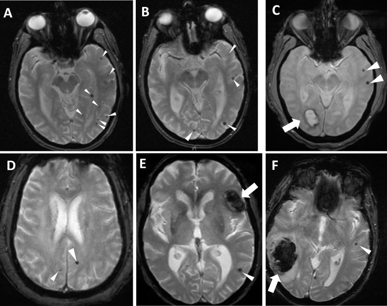
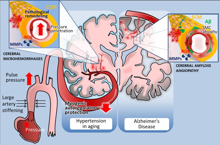
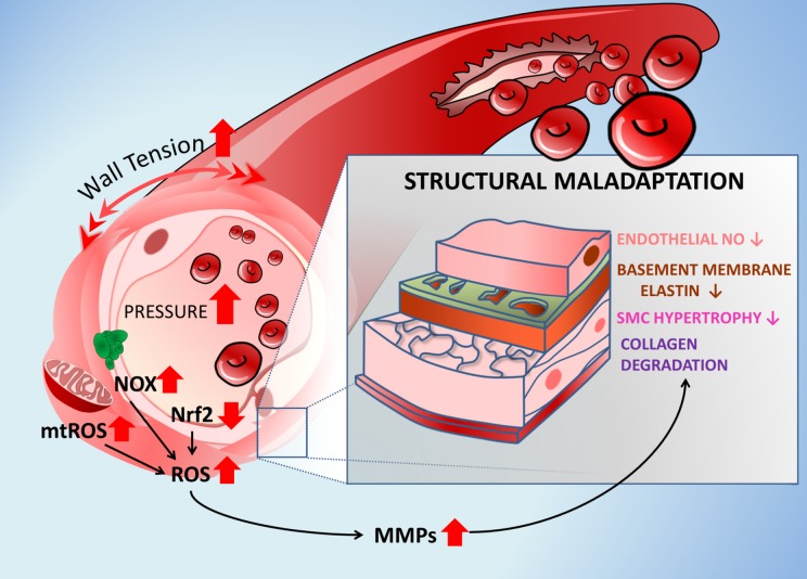
The increasing prevalence of multifocal cerebral microhemorrhages (CMHs, also known as "cerebral microbleeds") is a significant, newly recognized problem in the aging population of the Western world. CMHs are associated with rupture of small intracerebral vessels and are thought to progressively impair neuronal function, potentially contributing to cognitive decline, geriatric psychiatric syndromes, and gait disorders. Clinical studies show that aging and hypertension significantly increase prevalence of CMHs. CMHs are also now recognized by the National Institutes of Health as a major factor in Alzheimer's disease pathology. Moreover, the presence of CMHs is an independent risk factor for subsequent larger intracerebral hemorrhages. In this article, we review the epidemiology, detection, risk factors, clinical significance, and pathogenesis of CMHs. The potential age-related cellular mechanisms underlying the development of CMHs are discussed, with a focus on the structural determinants of microvascular fragility, age-related alterations in cerebrovascular adaptation to hypertension, the role of oxidative stress and matrix metalloproteinase activation, and the deleterious effects of arterial stiffening, increased pulse pressure, and impaired myogenic autoregulatory protection on the brain microvasculature. Finally, we examine potential treatments for the prevention of CMHs based on the proposed model of aging- and hypertension-dependent activation of the reactive oxygen species-matrix metalloproteinases axis, and we discuss critical questions to be addressed by future studies.

Keywords: cerebromicrovascular; cerebrovascular; stroke; transient ischemic attack; vascular aging; vascular cognitive impairment; vascular contributors to cognitive impairment and dementia.


Figures

Fig. 1.
Fig. 1.
Cerebral microhemorrhages visible on T2*-GRE MRI sequences in the axial plane (please see white arrowheads). All images were obtained on a 1.5 T field strength scanner (GE Medical Systems). A: 76-yr-old man with chronic untreated hypertension and mild cognitive impairment presenting with transient ischemic attack manifested as right hemiparesis. Imaging was negative for acute cerebral infarction. Although at least one lesion involves deeper brain regions, many of the cerebral microhemorrhages (CMHs) involve the gray-white matter junction. B: 82-yr-old woman with a diagnosis of probable Alzheimer's disease in the moderate stage of diabetes, coronary artery disease, and hypertension. Imaging was obtained as part of the outpatient assessment for memory loss and risk prediction for future risk of intracranial hemorrhage, as it was preceded by a concurrent new diagnosis of deep venous thrombosis. Several lobar CMHs are noted, suggesting amyloid angiopathy. C: 73-yr-old man with amnestic mild cognitive impairment, hypertension, coronary artery disease, and current smoking, presenting with right visual field deficit of unclear duration. Note the large occipital bleed (thick arrow) and additional two left hemispheric CMHs (arrowheads). D: 57-yr-old man with long-standing smoking, hypercholesterolemia, and untreated hypertension. He presented with transient ischemic attack manifested as hemisensory loss and dysarthria. E: 61-yr-old man presenting with new-onset mild aphasia and headache. He had a prior history of chronic anticoagulation for cardiac valvular disease, with therapeutic levels of warfarin upon presentation. Note the large intracerebral hemorrhage (thick arrow) and additional CMH (arrowhead), illustrating the potential association between CMHs and larger intraparenchymal bleeds. F: 86-yr-old woman with prior stroke, atrial fibrillation (not on anticoagulation), and hypertension presenting with acute stroke, treated with intravenous thrombolytics with subsequent development of intraparenchymal hemorrhage (thick arrow). Note the presence of CMH (thin arrow) in the contralateral hemisphere.
Fig. 2.
Fig. 2.
Distribution and pathogenesis of cerebral microhemorrhages (CMHs) associated with hypertension and Alzheimer’s disease in elderly patients. Left: in elderly patients CMHs associated with hypertensive vasculopathy typically affect the small perforating end-arteries located in the deep gray nuclei, brain stem, cerebellum, and deep white matter. The scheme highlights potential factors determining location and mechanisms involved in microvascular fragility. Accordingly, age-related large conduit artery stiffening increases pulsatile pressure, which can penetrate into the vulnerable portion of the microcirculation due to an impairment of myogenic autoregulatory protection in the proximal resistance arteries. The predilection of brain regions to hypertension-induced CMHs is determined by the branching pattern of penetrating arterioles. Increased pressure-induced vascular reactive oxygen species (ROS) production activates matrix metalloproteinases (MMPs), which degrade collagens and other components of the extracellular matrix (ECM), compromising the structural integrity of the cerebral microvasculature and promoting CMHs (inset). Right: in Alzheimer’s disease, CMHs develop in vessels affected by cerebral amyloid angiopathy (CAA). Location of the CMHs is determined by the predilection of brain regions for CAA pathology, which preferentially affects the small arteries and arterioles located in the cerebral cortex and at the junction of white and gray matter. The scheme depicts that deposition of Aβ in the vascular wall (green) promotes vascular smooth muscle cell atrophy and oxidative stress and exacerbates MMP-mediated degradation of the ECM, compromising the structural integrity of the vessels. The model predicts that because similar cellular mechanisms are involved in the pathogenesis of CMHs in both conditions, similar interventions should be effective for prevention as well.
Fig. 3.
Fig. 3.
Cellular mechanisms by which aging exacerbates development of CMHs. The scheme highlights the role of age-related exacerbation of pressure-induced vascular oxidative stress, MMP activation, and structural maladaptation to hypertension in pathogenesis of CMHs. Accordingly, the increased intraluminal pressure and consequential increases in wall tension activate NADPH oxidases and promote mitochondria-derived production of ROS (mtROS) in the aged vascular smooth muscle cells. Pressure-induced vascular oxidative stress is exacerbated in aging due to a homeostatic failure due to dysregulation of NRF2-mediated antioxidant response. Vascular oxidative stress is responsible for endothelial dysfunction and the increase in MMP activity, which is implicated in collagen degradation, smooth muscle cell atrophy, and degradation of elastic components of the basement membrane. These structural changes weaken the microvascular wall and increase vulnerability to the formation of CMHs. Arrows indicate the effects of aging.

References

    1. Alamowitch S, Plaisier E, Favrole P, Prost C, Chen Z, Van Agtmael T, Marro B, Ronco P. Cerebrovascular disease related to COL4A1 mutations in HANAC syndrome. Neurology 73: 1873–1882, 2009. doi:10.1212/WNL.0b013e3181c3fd12. - DOI - PMC - PubMed
    1. Altmann-Schneider I, Trompet S, de Craen AJ, van Es AC, Jukema JW, Stott DJ, Sattar N, Westendorp RG, van Buchem MA, van der Grond J. Cerebral microbleeds are predictive of mortality in the elderly. Stroke 42: 638–644, 2011. doi:10.1161/STROKEAHA.110.595611. - DOI - PubMed
    1. Aoki T, Kataoka H, Morimoto M, Nozaki K, Hashimoto N. Macrophage-derived matrix metalloproteinase-2 and -9 promote the progression of cerebral aneurysms in rats. Stroke 38: 162–169, 2007. doi:10.1161/01.STR.0000252129.18605.c8. - DOI - PubMed
    1. Arboleda-Velasquez JF, Manent J, Lee JH, Tikka S, Ospina C, Vanderburg CR, Frosch MP, Rodríguez-Falcón M, Villen J, Gygi S, Lopera F, Kalimo H, Moskowitz MA, Ayata C, Louvi A, Artavanis-Tsakonas S. Hypomorphic Notch 3 alleles link Notch signaling to ischemic cerebral small-vessel disease. Proc Natl Acad Sci USA 108: E128–E135, 2011. doi:10.1073/pnas.1101964108. - DOI - PMC - PubMed
    1. Armstrong M Jr, Strack GB. Recognition and documentation of strangulation crimes: a review. JAMA Otolaryngol Head Neck Surg 142: 891–897, 2016. doi:10.1001/jamaoto.2016.0293. - DOI - PubMed

MeSH terms

Substances

LinkOut - more resources