

tDCS-Induced Modulation of GABA Levels and Resting-State Functional Connectivity in Older Adults
- PMID: 28314813
- PMCID: PMC6596583
- DOI: 10.1523/JNEUROSCI.0079-17.2017
tDCS-Induced Modulation of GABA Levels and Resting-State Functional Connectivity in Older Adults
Abstract
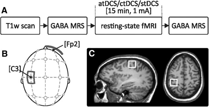
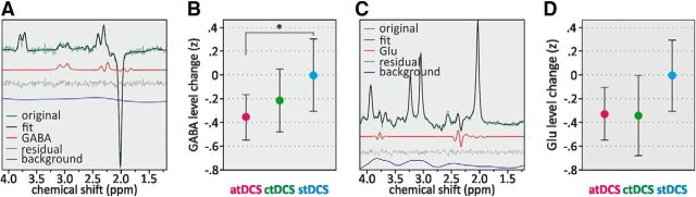
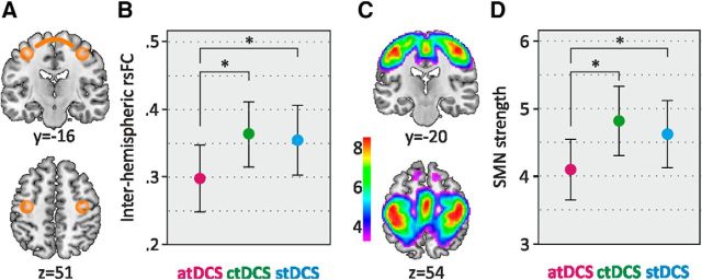
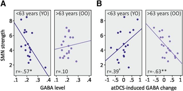
Transcranial direct current stimulation (tDCS) modulates human behavior, neuronal patterns, and metabolite concentrations, with exciting potential for neurorehabilitation. However, the understanding of tDCS-induced alterations on the neuronal level is incomplete, and conclusions from young adults, in whom the majority of studies have been conducted, cannot be easily transferred to older populations. Here, we investigated tDCS-induced effects in older adults (N = 48; age range, 50-79 years) using magnetic resonance spectroscopy to quantify GABA levels as well as resting-state functional magnetic resonance imaging to assess sensorimotor network strength and interhemispheric connectivity. In a randomized, counterbalanced, crossover design, we applied anodal tDCS (atDCS), cathodal tDCS (ctDCS), and sham tDCS (stDCS) over the left sensorimotor region. We observed a significant reduction of GABA levels after atDCS compared with stDCS, reflecting the preserved neuromodulatory effect of atDCS in older adults. Moreover, resting-state functional coupling was decreased during atDCS compared with stDCS, most likely indicating augmented efficiency in brain network functioning. Increased levels of interhemispheric connectivity with age were diminished by atDCS, suggesting stimulation-induced functional decoupling. Further, the magnitude of atDCS-induced local plasticity was related to baseline functional network strength. Our findings provide novel insight into the neuronal correlates underlying tDCS-induced neuronal plasticity in older adults and thus might help to develop tDCS interventions tailored to the aging brain.SIGNIFICANCE STATEMENT Transcranial direct current stimulation (tDCS) modulates human behavior, neuronal patterns, and metabolite concentrations, with exciting potential for neurorehabilitation. However, the understanding of tDCS-induced alterations on the neuronal level is incomplete, and conclusions from young adults cannot be easily transferred to older populations. We used a systematic multimodal imaging approach to investigate the neurophysiological effects of tDCS in older adults and found stimulation-induced effects on GABA levels, reflecting augmented local plasticity and functional connectivity, suggesting modulation of network efficiency. Our findings may help to reconcile some of the recent reports on the variability of tDCS-induced effects, not only implicating age as a crucial modulating factor, but detailing its specific impact on the functionality of neural networks.
Keywords: aging; magnetic resonance spectroscopy; plasticity; sensorimotor network; transcranial electrical stimulation.
Copyright © 2017 the authors 0270-6474/17/374065-09$15.00/0.
Figures
References
Publication types
MeSH terms
Substances
LinkOut - more resources
Full Text Sources
Other Literature Sources
Medical