

Live-Cell Imaging of Protease Activity: Assays to Screen Therapeutic Approaches
- PMID: 28315254
- PMCID: PMC5500169
- DOI: 10.1007/978-1-4939-6850-3_16
Live-Cell Imaging of Protease Activity: Assays to Screen Therapeutic Approaches
Abstract
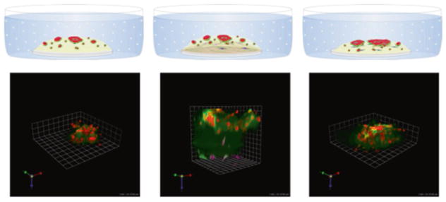
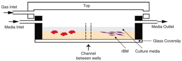
Methodologies to image and quantify the activity of proteolytic enzymes have been developed in an effort to identify protease-related druggable pathways that are involved in malignant progression of cancer. Our laboratory has pioneered techniques for functional live-cell imaging of protease activity in pathomimetic avatars for breast cancer. We analyze proteolysis in the context of proliferation and formation of structures by tumor cells in 3-D cultures over time (4D). In order to recapitulate the cellular composition and architecture of tumors in the pathomimetic avatars, we include other tumor-associated cells (e.g., fibroblasts, myoepithelial cells, microvascular endothelial cells). We also model noncellular aspects of the tumor microenvironment such as acidic pericellular pH. Use of pathomimetic avatars in concert with various types of imaging probes has allowed us to image, quantify, and follow the dynamics of proteolysis in the tumor microenvironment and to test interventions that impact directly or indirectly on proteolytic pathways. To facilitate use of the pathomimetic avatars for screening of therapeutic modalities, we have designed and fabricated custom 3D culture chambers with multiple wells that are either individual or connected by a channel to allow cells to migrate between wells. Optical glass microscope slides underneath an acrylic plate allow the cultures to be imaged with an inverted microscope. Fluid ports in the acrylic plate are at a level above the 3D cultures to allow introduction of culture media and test agents such as drugs into the wells and the harvesting of media conditioned by the cultures for immunochemical and biochemical analyses. We are using the pathomimetic avatars to identify druggable pathways, screen drug and natural product libraries and accelerate entry of validated drugs or natural products into clinical trials.
Keywords: Confocal microscopy; Fluorescent imaging; High-content imaging; Proteolysis; Screening assays.
Figures
References
-
- Edwards D, Høyer-Hansen G, Blasi F, Sloane BF. The cancer degradome: proteases and cancer biology. Springer-Verlag; New York: 2008.
-
- Sloane BF, List K, Fingleton B, Matrisian L. Proteases in cancer—significance for invasion and metastasis. In: Brix K, Stoecker W, editors. Proteases—structure and function. Springer-Verlag Wien; Vienna: 2013. pp. 491–550.
-
- Lopez-Otin C, Matrisian LM. Emerging roles of proteases in tumour suppression. Nat Rev Cancer. 2007;7(10):800–808. - PubMed
-
- Schwartz DR, Moin K, Yao B, Matrisian LM, Coussens LM, Bugge TH, Fingleton B, Acuff HB, Sinnamon M, Nassar H, Platts AE, Krawetz SA, Linebaugh BE, Sloane BF. Hu/Mu ProtIn oligonucleotide microarray: dual-species array for profiling protease and protease inhibitor gene expression in tumors and their microenvironment. Mol Cancer Res. 2007;5(5):443–454. - PubMed
Publication types
MeSH terms
Substances
Grants and funding
LinkOut - more resources
Full Text Sources
Other Literature Sources
