. 2017 Apr 4;89(7):3819-3823.
doi: 10.1021/acs.analchem.7b00470. Epub 2017 Mar 20.

Evaluation of Nanobody Conjugates and Protein Fusions as Bioanalytical Reagents

Affiliations

Evaluation of Nanobody Conjugates and Protein Fusions as Bioanalytical Reagents

Virginia J Bruce et al. Anal Chem. .

Abstract

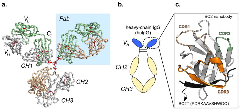
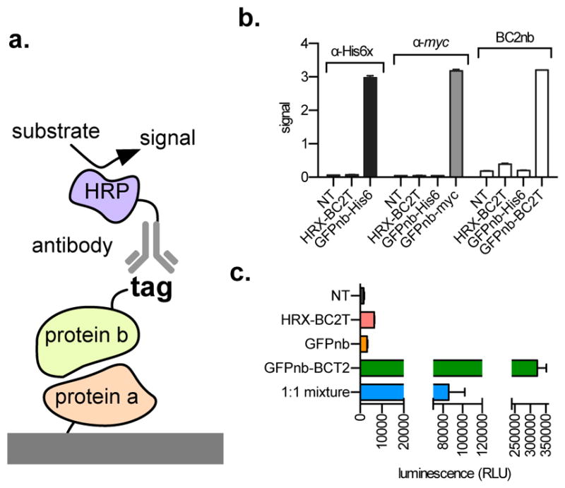
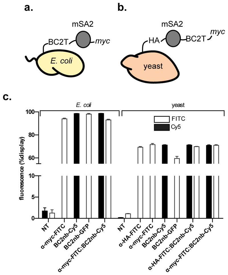
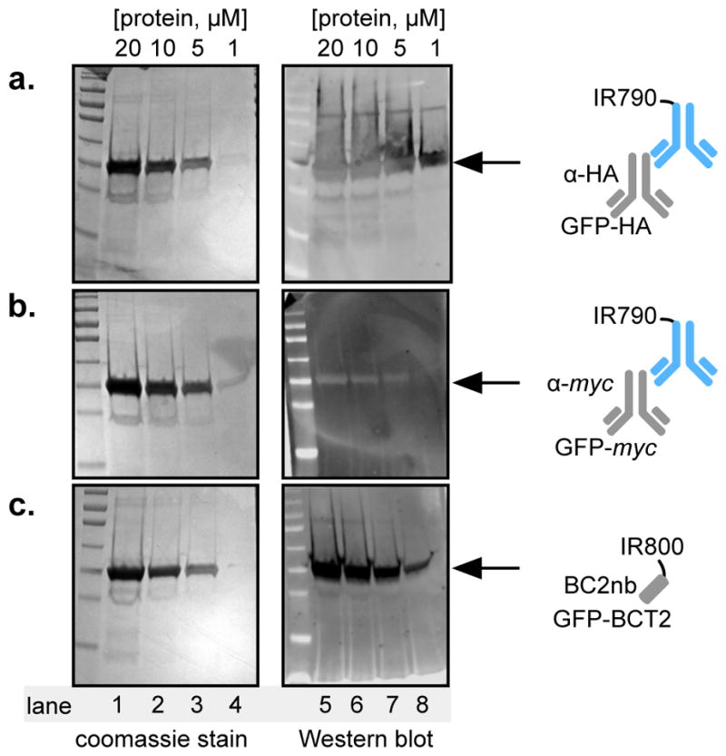
Enzyme-linked immunosorbent assay (ELISA), flow cytometry, and Western blot are common bioanalytical techniques. Successful execution traditionally requires the use of one or more commercially available antibody-small-molecule dyes or antibody-reporter protein conjugates that recognize relatively short peptide tags (<15 amino acids). However, the size of antibodies and their molecular complexity (by virtue of post-translational disulfide formation and glycosylation) typically require either expression in mammalian cells or purification from immunized mammals. The preparation and purification of chemical dye- or reporter protein-antibody conjugates is often complicated and expensive and not commonplace in academic laboratories. In response, researchers have developed comparatively simpler protein scaffolds for macromolecular recognition, which can be expressed with relative ease in E. coli and can be evolved to bind virtually any target. Nanobodies, a minimalist scaffold generated from camelid-derived heavy-chain IgGs, are one such example. A multitude of nanobodies have been evolved to recognize a diverse array of targets, including a short peptide. Here, this peptide tag (termed BC2T) and BC2 nanobody-dye conjugates or reporter protein fusions are evaluated in ELISA, flow cytometry, and Western blot experiments and compared to analogous experiments using commercially available antibody-conjugate/peptide tag pairs. Collectively, the utility and practicality of nanobody-based reagents in bioanalytical chemistry is demonstrated.


Figures

Figure 1
Figure 1
(a) Structure of IgG. Disulfide bonds are highlighted in red. Constant heavy-chain region 1 (CH1); constant light-chain (CL); variable light-chain (VL); variable heavy-chain (VH), and fragment antigen-binding (Fab) regions are highlighted with a blue background (PDB: 1IGY). (b) Architecture of a heavy-chain IgG (hcIgG), consisting of two heavy chains (CH3, CH2, VH) connected by disulfide bonds in the hinge region. The “nanobody” subunit is circled. (c) Structure of the recently reported nanobody BC2, bound to its peptide tag (BC2T, PDB: 5IVN). This complex was originally reported in Nature Scientific Reports 6, 19211 (2016).
Figure 2
Figure 2
(a) Scheme of an Enzyme-Linked Immunosorbent Assay (ELISA). (b) ELISA data: immobilized GFP is treated with buffer (NT), and either HRX-BCT2, GFPnb-His6, GFPnb-myc, or GFPnb-BC2T, then either anti-His6-HRP, anti-myc-HRP, or the BC2nb-HRP conjugate, and HRP substrate. Signal is the observed absorbance at 655 nm. (c) ELISA data: GFP was immobilized onto streptavidin coated plates, then treated with buffer (NT, black), HRX-BC2T (red), GFP nanobody (GFPnb, orange), GFPnb-BC2T (green), or a 1:1 mixture of GFP nanobody and GFPnb-BC2T (blue), followed by nLuc substrate. All experiments were performed in triplicate. Error bars represent standard deviation of three experiments. α = anti; NT = no treatment. RLU = relative luminescence units.
Figure 3
Figure 3
(a) Representation of E. coli engineered for flow cytometry experiments. (b) Representation of yeast engineered for flow cytometry experiments. (c) Flow cytometry detection of displayed monomeric streptavidin (mSA2) on the surface of E. coli or yeast, as determined by commercially available antibody α-myc-FITC, or nanobody reagents BC2nb-Cy5, or BC2nb-GFP (for E. coli), or commercially available antibodies α-myc-FITC, or α-HA-FITC, or nanobody reagents BC2nb-Cy5, or BC2nb-GFP (for yeast). All experiments were performed in triplicate. Error bars represent standard deviation of three experiments. α = anti; NT = no treatment.
Figure 4
Figure 4
(a–c, left gel) Coomassie stained polyacrylamide gels following loading with 20, 10, 5, or 1 μM GFP-HA, GFP-myc, or GFP-BCT2, and electrophoresis. (a–c, right gel) Western blot data for the GFP-HA/anti-HA; GFP-myc/anti-myc, or; GFP-BC2T/BC2nb pairs, respectively. α = anti.

References

    1. Byrne B, Stack E, Gilmartin N, O’Kennedy R. Sensors (Basel) 2009;9:4407–4445. - PMC - PubMed
    1. Holford TR, Davis F, Higson SP. Biosens Bioelectron. 2012;34:12–24. - PubMed
    1. Liu ZQ, Mahmood T, Yang PC. N Am J Med Sci. 2014;6:160. - PMC - PubMed
    1. Bull World Health Organ. 1976;54:129–139. - PMC - PubMed
    1. Laerum OD, Farsund T. Cytometry. 1981;2:1–13. - PubMed

Publication types

MeSH terms

LinkOut - more resources