

Case Reports
doi: 10.1155/2017/3421832.
Epub 2017 Feb 20.
Retiform Sertoli-Leydig Cell Tumor in a 38-Year-Old Woman: A Case Report, Retrospective Review, and Review of Current Literature
Affiliations
- PMID: 28316852
- PMCID: PMC5337871
- DOI: 10.1155/2017/3421832
Item in Clipboard
Case Reports
Retiform Sertoli-Leydig Cell Tumor in a 38-Year-Old Woman: A Case Report, Retrospective Review, and Review of Current Literature
Case Rep Pathol.
2017.
Abstract
Ovarian sex cord-stromal tumors arise from the stromal cells that surround and support the oocytes. Sertoli-Leydig cell tumors belong to this category of ovarian neoplasms. We present the case of a 38-year-old woman who was found to have a right ovarian mass. The mass was resected and diagnosed as Stage I Sertoli-Leydig cell tumor, retiform variant, following histopathologic and immunohistochemical examination. This case is unusual given the rarity of the retiform variant of Sertoli-Leydig cell tumor and the atypically older age of 38 years at presentation.
Conflict of interest statement
The authors declare that they have no competing interest.
Figures

Gross image of right ovarian solid and multicystic mass which measured 27.0 × 17.0 × 5.0 cm. The capsular surface showed a 4.0 × 2.0 cm area of disruption (blue arrow). A 4.3 × 3.0 × 0.6 cm solid area was also noted (red arrow).

Sections from the tumor show networks of anastomosing slit-like to cystic spaces lined by cuboidal Sertoli cells, resembling the rete testis. Some of the retiform tubules are denoted by blue arrows (H&E, 100x).

Another section of the tumor demonstrates sheets of Sertoli cells. Within the sheets of Sertoli cells, areas of small tubular formation can be seen. Clusters of Leydig cells are also noted (blue arrows) (H&E, 100x).

A section of the tumor shows the solid component of the tumor composed of Sertoli cells. Small tubular formation is noted in the solid component. Cuboidal Sertoli cells line the slit-like spaces (H&E, 200x).

Image of the tumor demonstrating the merging of the solid component composed of Sertoli cells with the Sertoli cells lining the slit-like spaces (H&E, 400x).

Immunohistochemical profile of the tumor for pancytokeratin, beta-catenin, calretinin, and inhibin (20x).
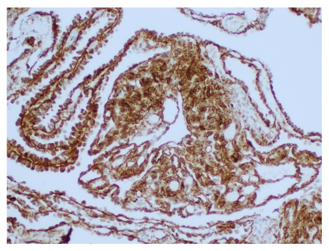
Nuclear and cytoplasmic beta-catenin positivity in retiform areas and focal solid areas composed of sheets of Sertoli cells (200x).

Beta-catenin with nuclear and cytoplasmic positivity in retiform areas (400x).

Beta-catenin with nuclear and cytoplasmic positivity in Leydig cells (400x).
References
-
- World Health Organization. WHO Classification of Tumours of Female Reproductive Organs. 4th. Vol. 6. Lyon, France: International Agency for Research on Cancer; 2014. Edited by R. J. Kurman, M. L. Carcangiu, C. S. Herrington, and R. H. Young.
-
- Mills S. E., Greenson J. K., Hornick J. L. Sternberg's Diagnostic Surgical Pathology. 6th. Vol. 2. Philadelphia, Pa, USA: Wolters Kluwer; 2015. Ovarian epithelial-stromal tumors; pp. 2584–2587.
-
- Adouni O., Kourda N., Aloui R., Zermani R., Abdellatif C., Ben Jilani S. [Retiform Sertoli-Leydig tumor with heterologous mucinous differentiation] La Tunisie médicale. 2012;90(2):187–189. - PubMed
Publication types
LinkOut - more resources
Full Text Sources
Other Literature Sources
