. 2017 Mar 20:8:14179.
doi: 10.1038/ncomms14179.

Polymeric mechanical amplifiers of immune cytokine-mediated apoptosis

Affiliations

Polymeric mechanical amplifiers of immune cytokine-mediated apoptosis

Michael J Mitchell et al. Nat Commun. .

Abstract

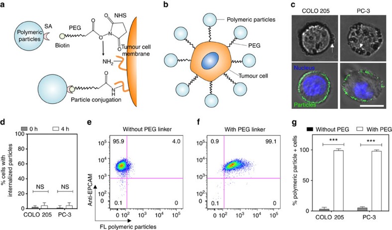
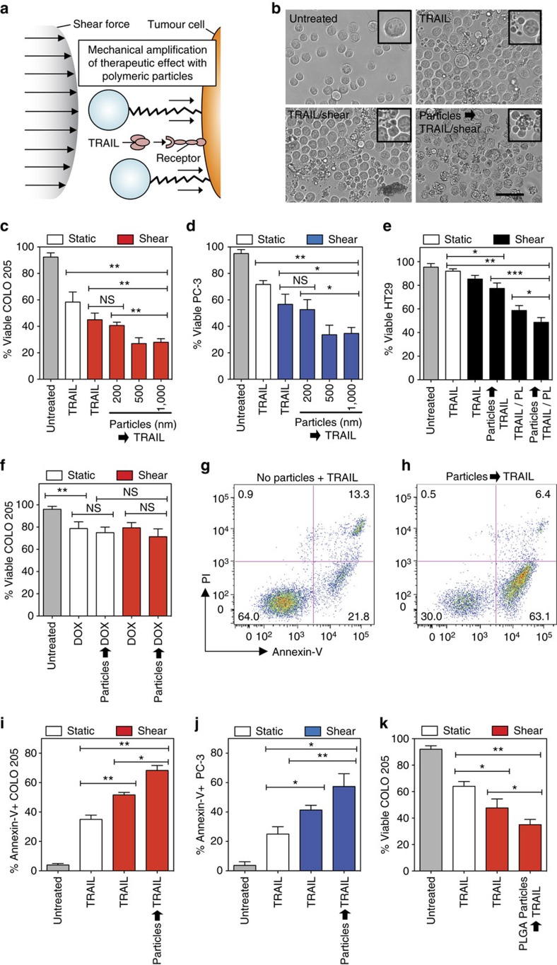
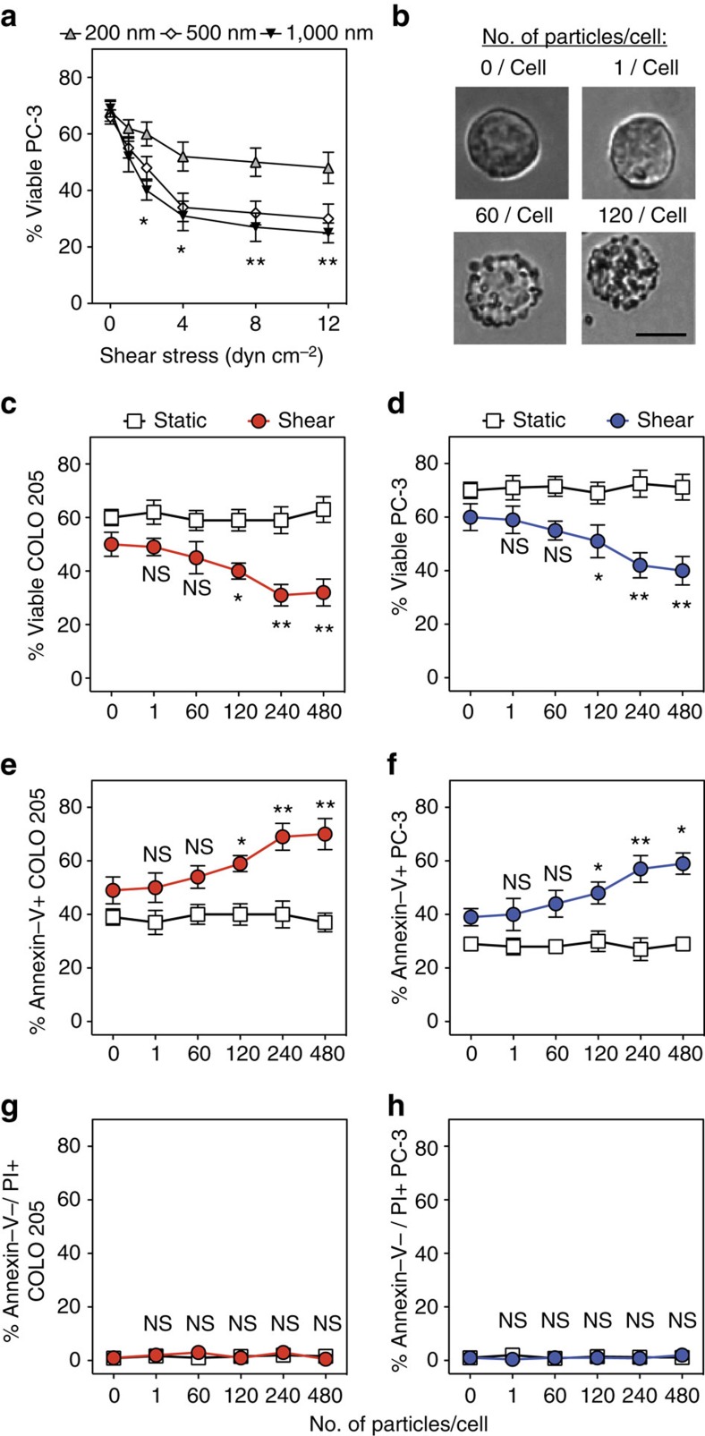
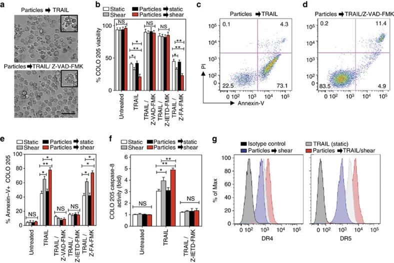
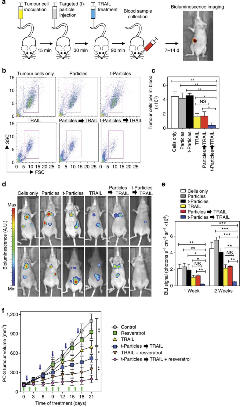
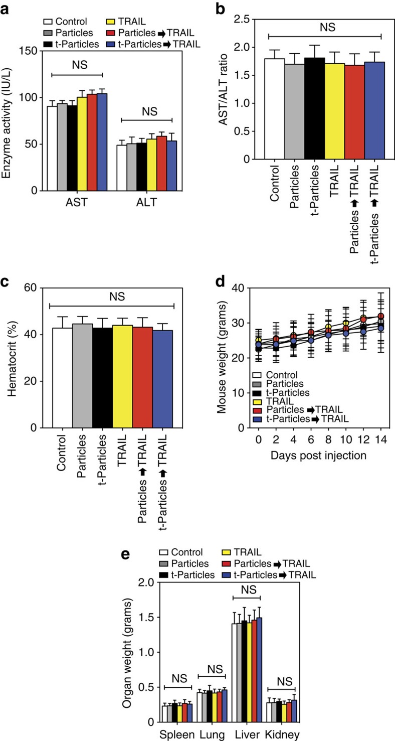
Physical forces affect tumour growth, progression and metastasis. Here, we develop polymeric mechanical amplifiers that exploit in vitro and in vivo physical forces to increase immune cytokine-mediated tumour cell apoptosis. Mechanical amplifiers, consisting of biodegradable polymeric particles tethered to the tumour cell surface via polyethylene glycol linkers, increase the apoptotic effect of an immune cytokine on tumour cells under fluid shear exposure by as much as 50% compared with treatment under static conditions. We show that targeted polymeric particles delivered to tumour cells in vivo amplify the apoptotic effect of a subsequent treatment of immune cytokine, reduce circulating tumour cells in blood and overall tumour cell burden by over 90% and reduce solid tumour growth in combination with the antioxidant resveratrol. The work introduces a potentially new application for a broad range of micro- and nanoparticles to maximize receptor-mediated signalling and function in the presence of physical forces.


Conflict of interest statement

The authors declare no competing financial interests.

Figures

Figure 1
Figure 1. Functionalization of the tumour cell surface with polymeric particles.
(a,b) NHS–PEG–biotin linkers (a) were used to conjugate a range of streptavidin-functionalized polymeric particles to the tumour cell surface (b). (c) Brightfield (top) and confocal (bottom) micrographs of polymeric polystyrene (PS) particles conjugated to the surface of colon (COLO 205; left) and prostate (PC-3; right) tumour cell lines. The 500 nm diameter PS particles bound to tumour cells in brightfield micrographs. Brightfield micrographs show 500 nm diameter PS particles bound to tumour cells. The 200 nm diameter PS particles bound to tumour cells in confocal micrographs. Confocal micrographs shown 200 nm diameter PS particles bound to tumour cells. Green indicates polymeric PS particles and blue indicates nucleus. Scale bar, 10 μm. (d) Percentage of tumour cells with internalized fluorescent PS particles, immediately after and 4 h post functionalization. Internalized fluorescent PS particles quantified using a Trypan blue fluorescence quenching assay and confocal microscopy. N=5 per treatment. (e,f) Flow cytometry plots of epithelial cell adhesion molecule (EpCAM)+ tumour cells functionalized with fluorescent PS particles in the absence (e) and presence (f) of a PEG linker. N=5 per treatment. (g) Percentage of fluorescent PS particle+ tumour cells after functionalization with a PEG linker. Data are reported as the mean±s.e. Different treatment groups were compared for statistical significance using Student's two-tailed t-test. N=5 per treatment. NHS, N-hydroxysuccinimide; NS, not significant; SA, streptavidin. ***P<0.001.
Figure 2
Figure 2. Polymeric particles conjugated to tumour cell surface amplify TRAIL-mediated apoptosis in the presence of fluid shear stress.
(a) Schematic of polymeric particles acting as mechanical amplifiers by increasing TRAIL-mediated tumour cell apoptosis in presence of a fluid shear force. (b) Brightfield micrographs of particle-functionalized COLO 205 tumour cells after 30 min of exposure to various treatment conditions. TRAIL-treated samples incubated with 0.1 μg ml−1 TRAIL for 30 min. Sheared samples were exposed to a fluid shear stress of 4.0 dyn cm−2. Tumour cells were treated with 240 PS (500 nm diameter) particles per tumour cell before exposure to TRAIL and fluid shear stress. Insets denote viable and apoptotic tumour cells after treatment conditions. Scale bar, 30 μm. (c,d) Viability of particle-functionalized COLO 205 (c) and PC-3 (d) tumour cells after treatment with TRAIL (0.1 μg ml−1) in the presence of fluid shear stress. N=4 per treatment. (e) Viability of particle-functionalized, TRAIL-resistant HT29 tumour cells after treatment with TRAIL (0.1 μg ml−1) in the presence of fluid shear stress for 6 h. For combination therapies, cells were treated with 15 μM piperlongumine (PL) in addition to TRAIL (0.1 μg ml−1). N=4 per treatment. (f) Viability of particle-functionalized COLO 205 tumour cells after treatment with doxorubicin (DOX; concentration: 20 μM) in the presence of fluid shear stress. N=4 per treatment. Particle diameter: 500 nm. (g,h) Annexin-V/propidium iodide (PI) flow cytometry plots of nonfunctionalized (g) and particle (500 nm)-functionalized (h) PC-3 tumour cells after treatment with TRAIL (0.1 μg ml−1) in the presence of static conditions and fluid shear stress, respectively. Particle diameter: 500 nm. N=5 per treatment. (i,j) Percentage of annexin-V+, particle-functionalized COLO 205 (i) and PC-3 (j) tumour cells after treatment with TRAIL (0.1 μg ml−1) in the presence of fluid shear stress. N=5 per treatment. (k) Viability of biodegradable PLGA particle-functionalized COLO 205 tumour cells after treatment with TRAIL (0.1 μg ml−1) in the presence of fluid shear stress. Data are reported as the mean±s.e. Different treatment groups were compared for statistical significance using Student's two-tailed t-test. Particle diameter: 500 nm. N=5 per treatment. *P<0.05, **P<0.01 and ***P<0.001. NS, not significant.
Figure 3
Figure 3. Increased shear stress, particle diameter and number of particles conjugated to tumour cell surface enhance TRAIL-mediated apoptosis.
(a) Viability of particle-functionalized PC-3 tumour cells treated with TRAIL in the presence of a range of shear forces (1.0–12.0 dyn cm−2) across a range of particle sizes (diameter: 200–1,000 nm). (b) COLO 205 tumour cells treated with 0–120 PS particles per cell in suspension. Scale bar, 10 μm. (c,d) Viability of particle-functionalized COLO 205 (c) and PC-3 tumour cells (d) treated with TRAIL in the presence of fluid shear stress. N=5 per treatment. (e,f) Percentage of annexin-V+ particle-functionalized COLO 205 (e) and PC-3 cells (f) treated with TRAIL in the presence of fluid shear stress. N=5 per treatment. (g,h) Percentage of annexin-V-/PI+ particle-functionalized COLO 205 (g) and PC-3 cells (h) treated with TRAIL in the presence of fluid shear stress (shear stress: 4.0 dyn cm−2). All tumour cells were incubated with 0–480 PS particles (500 nm diameter) per cell before all treatments. N=5 per treatment. TRAIL concentration: 0.1 μg ml−1 for all TRAIL-treated samples. Shear stress: 4.0 dyn cm−2 for all samples exposed to shear. Data are reported as mean±s.e. Different treatment groups were compared for statistical significance using Student's two-tailed t-test. NS, not significant. *P<0.05 and **P<0.01.
Figure 4
Figure 4. Amplification of TRAIL apoptotic effect via polymeric particles conjugation is caspase dependent and increases death receptor expression.
(a) Brightfield micrographs of particle (diameter: 500 nm)-functionalized COLO 205 tumour cells treated with 0.1 μg ml−1 TRAIL under fluid shear stress exposure (shear stress: 4.0 dyn cm−2) for 1 h in the absence and presence of 50 μM pan caspase inhibitor Z-VAD-FMK. (b) Viability of particle-functionalized COLO 205 tumour cells treated with 0.1 μg ml−1 TRAIL under fluid shear stress exposure for 1 h in the presence of 50 μM pan caspase inhibitor Z-VAD-FMK, 50 μM caspase-8 inhibitor Z-IETD-FMK or 50 μM caspase negative control inhibitor Z-FA-FMK. N=4 for all treatments. (c,d) Annexin-V/propidium iodide (PI) flow cytometry plots of particle-functionalized COLO 205 tumour cells treated with 0.1 μg ml−1 TRAIL in the presence of a fluid shear stress for 1 h without and with Z-VAD-FMK treatment. N=4 for all treatments. (e) Annexin-V quantification of particle-functionalized COLO 205 tumour cells treated with 0.1 μg ml−1 TRAIL under fluid shear stress exposure for 1 h in the presence of 50 μM pan caspase inhibitor Z-VAD-FMK, 50 μM caspase-8 inhibitor Z-IETD-FMK or 50 μM caspase negative control inhibitor Z-FA-FMK. N=4 for all treatments. (f) Caspase-8 activity of particle-functionalized COLO 205 tumour cells after treatment with TRAIL (0.1 μg ml−1) in the presence and absence of fluid shear stress for 1 h. N=4 for all treatments. (g) TRAIL death receptor (DR) 4 and 5 expression after treatment of particle-functionalized COLO 205 tumour cells with TRAIL (0.1 μg ml−1) in the presence and absence of fluid shear stress for 1 h. N=5 for all treatments. Data are reported as mean±s.e. Different treatment groups were compared for statistical significance using Student's two-tailed t-test for two conditions and one-way analysis of variance (ANOVA) for multiple comparisons. Percent of max represents the number of events normalized according to FlowJo algorithms. *P<0.05 and **P<0.01. NS, not significant.
Figure 5
Figure 5. Polymeric particles targeted to tumor cell surface amplify immune cytokinemediated apoptosis in vivo.
(a) Schematic of epithelial cell adhesion molecule (EpCAM)-targeted particle delivery to COLO 205 tumour cells in nude (nu/nu) mice in vivo, followed by treatment with TRAIL. Mice were inoculated with COLO 205 tumour cells via tail vein injection (2 × 10⁶ cells), followed by injection of nontargeted and EpCAM-targeted PLGA particles (500 nm diameter; ∼500 particles per tumour cell) 15 min post tumour cell injection. At 30 min post particle injection, mice were treated with TRAIL (0.1 μg ml−1 plasma concentration). Tumour cells in blood were collected via submandibular bleed 90 min post TRAIL injection. Tumour cells were detected in vivo via whole-body bioluminescent imaging (BLI) at 7 and 14 days post injection. (b) Representative flow cytometry plots of GFP+ COLO 205 tumour cells removed after delivery of nontargeted particles (Particles) and EpCAM-targeted particles (t-Particles) followed by TRAIL. FSC, forward scatter; SSC, side scatter. (c) Number of viable GFP+ COLO 205 tumour cells per ml mouse blood 90 min post TRAIL treatment of tumour cells in vivo under various conditions. Cells only denotes mice treated with tumour cells followed by PBS via tail vein injection. N=5 mice for all treatments. (d) Representative whole-body BLI images of COLO 205 tumour cells in mice 7 days post injection of particles and targeted particles followed by TRAIL. (e) COLO 205 BLI signals in mice 7 and 14 days post injection of COLO 205 tumour cells under various conditions. N=5 mice for all treatments. (f) PC-3 tumour growth curves after intravenous injections of targeted particles (40 mg kg−1) followed by TRAIL (15 mg kg−1) 3 h post particle injection. For combination therapies, tumour-bearing nu/nu mice were also treated with the TRAIL-sensitizer resveratrol (30 mg kg−1). After tumour formation (100 mm3), mice began treatment regimen and tumour volume was measured every 3 days. Blue arrows indicate days where mice were treated with targeted particles, followed 3 h later by TRAIL treatment. Green arrows indicate days where mice were treated with resveratrol via oral gavage. N=5 mice for all treatments. Data are reported as the mean±s.e. Different treatment groups were compared for statistical significance using Student's two-tailed t-test for two conditions and one-way analysis of variance (ANOVA) for multiple comparisons. *P<0.05, **P<0.01 and ***P<0.001. NS, not significant.
Figure 6
Figure 6. Delivery of nontargeted particles (Particles) and EpCAM-targeted PLGA particles (t-Particles) followed by TRAIL therapeutic does not affect non-target cells and tissues in vivo.
(a) Serum levels of aspartate aminotransferase (AST) and alanine aminotransferase (ALT) liver enzymes in mice from controls and different treatment groups at the end of the 2-week study. (b) AST/ALT ratio in serum of mice from controls and different treatment groups at the end of the 2-week study. AST/ALT >2 indicates liver toxicity. (c) Haematocrit levels of mice from controls and different treatment groups. Blood was drawn before killing of mice. (d) Weight of mice 0–2 weeks post injection of TRAIL and particle-functionalized tumour cells. (e) Weight of excised organs post-mortem. Data are reported as mean±s.e. Different treatment groups were compared for statistical significance using a one-way analysis of variance (ANOVA) for multiple comparisons. NS, not significant. N=5 mice for all treatments.

Similar articles

Cited by

References

    1. Jordan J. D., Landau E. M. & Iyengar R. Signaling networks: the origins of cellular multitasking. Cell 103, 193–200 (2000). - PMC - PubMed
    1. Stevens M. M. & George J. H. Exploring and engineering the cell surface interface. Science 310, 1135–1138 (2005). - PubMed
    1. Bettinger C. J. & Langer R. Engineering substrate topography at the micro- and nanoscale to control cell function. Angew. Chem. 48, 5406–5415 (2009). - PMC - PubMed
    1. Mannix R. J. et al.. Nanomagnetic actuation of receptor-mediated signal transduction. Nat. Nanotechnol. 3, 36–40 (2007). - PubMed
    1. Lee J.-H. et al.. Artificial control of cell signaling and growth by magnetic nanoparticles. Angew. Chem. Int. Ed. 49, 5698–5702 (2010). - PubMed

Publication types

LinkOut - more resources