. 2017 Mar 20:7:44832.
doi: 10.1038/srep44832.

Structural and functional studies of a noncanonical Dicer from Entamoeba histolytica

Affiliations

Structural and functional studies of a noncanonical Dicer from Entamoeba histolytica

Xiang Yu et al. Sci Rep. .

Abstract

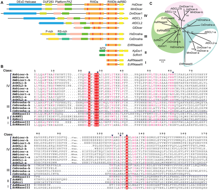
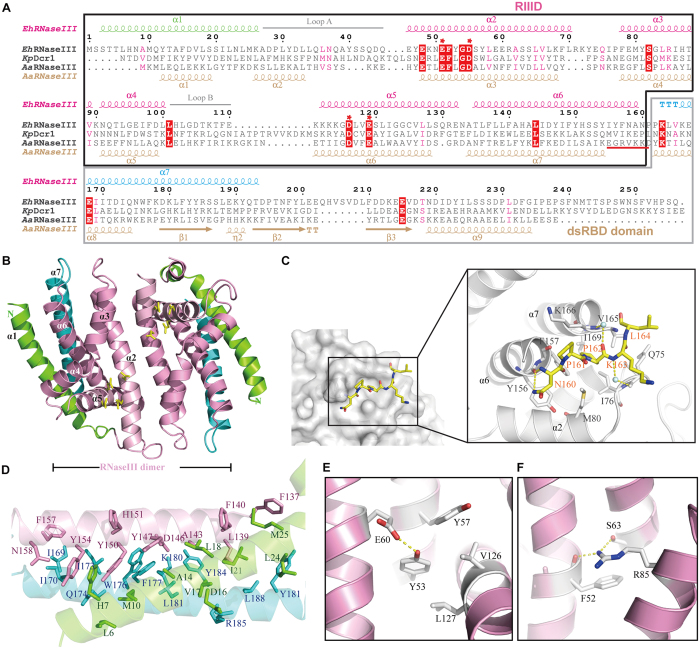
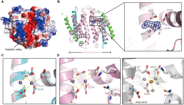
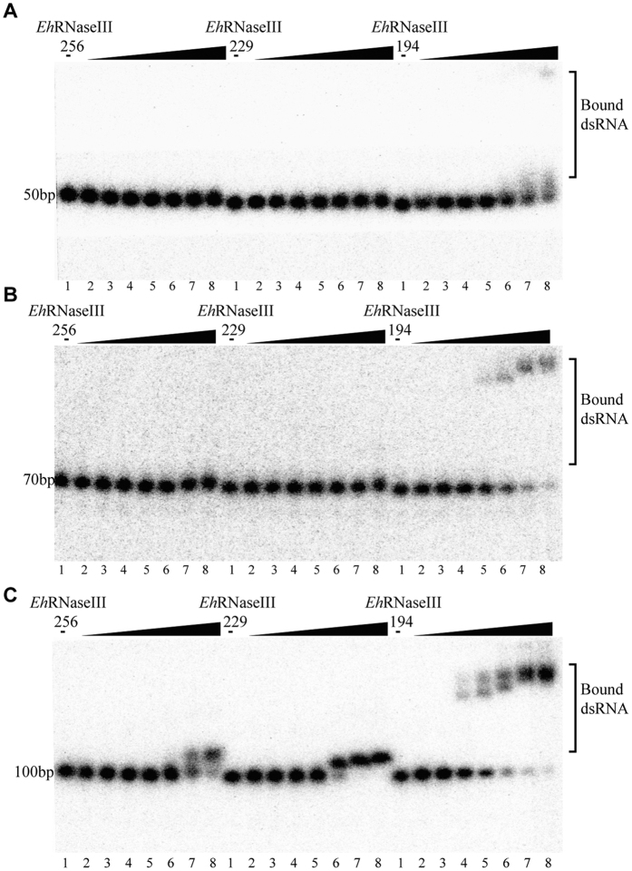
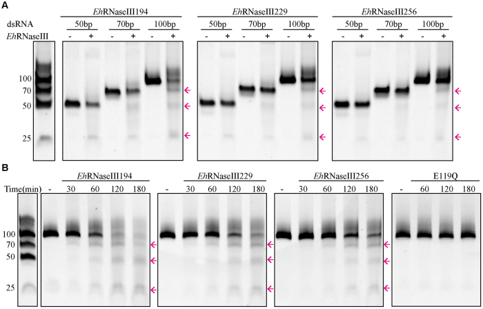
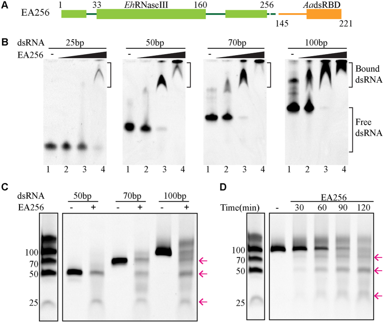
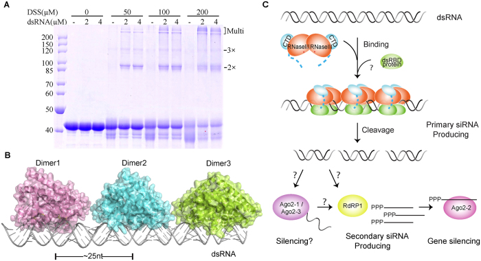
RNaseIII proteins are dsRNA-specific endonucleases involved in many important biological processes, such as small RNA processing and maturation in eukaryotes. Various small RNAs have been identified in a protozoan parasite Entamoeba histolytica. EhRNaseIII is the only RNaseIII endonuclease domain (RIIID)-containing protein in E. histolytica. Here, we present three crystal structures that reveal several unique structural features of EhRNaseIII, especially the interactions between the two helixes (α1 and α7) flanking the RIIID core domain. Structure and sequence analysis indicate that EhRNaseIII is a noncanonical Dicer and it lacks a dsRBD in the C-terminal region (CTR). In vitro studies suggest that EhRNaseIII prefers to bind and cleave longer dsRNAs, generating products around 25 nucleotides in length. Truncation of the CTR or attaching the dsRBD of Aquifex aeolicus RNaseIII can enhance the binding and cleavage activities of EhRNaseIII. In combination with in vitro crosslinking assay, our results suggested that EhRNaseIII functions in a cooperative mode. We speculate that some partner proteins may exist in E. histolytica and regulates the activity of EhRNaseIII through interaction with its CTR. Our studies support that EhRNaseIII plays an important role in producing small RNAs in E. histolytica.


Conflict of interest statement

The authors declare no competing financial interests.

Figures

Figure 1
Figure 1. EhRNaseIII is a non-canonical Dicer.
(A) Domain architectures of representative RNaseIIIs; tandem RIIID domains of Drosha and Dicer are designated as RIIIDa and RIIIDb, respectively. Hs, Homo sapiens; Mm, Mus musculus; Dm, Drosophila melanogaster; At, Arabidopsis thaliana; Gi, Giardia intestinalis; Ce, Caenorhabditis elegans; Sc, Saccharomyces cerevisiae; Kp, Kluyveromyces polysporus; Eh, Entamoeba histolytica; Aa, Aquifex aeolicus; Ec, Escherichia coli. (B) Sequence alignment of RIIID domains from each class of RNase III enzyme. HsDicer1, GI: 152012889; HsDrosha, GI: 20139357; MmDicer, GI: 257051057; DmDicer1, GI: 7300916; AtDCL1, GI: 34922211; AtDCL2, GI: 332640405; CeDrosha, GI: 20141625; ScRNT1, GI: 618855177; KpDcr1, GI: 342351115; EhRNaseIII, GI: 56467134; AaRNaseIII, GI: 160877684; EcRNaseIII, GI: 485668531. Conserved catalytic residues are indicated by asterisks, newly identified catalytic residues are labeled with an inverted triangle. (C) Maximum-likelihood tree of RIIID domains from RNaseIII families. RIIIDa and RIIIDb domains of class IV RNaseIIIs are colored white and pink, respectively. RIIIDa and RIIIDb domains of class III RNaseIIIs are colored cyan and green, respectively. Class II RNaseIIIs are also colored green. Class I RNaseIIIs are colored orange. Bs, Bacillus subtilis.
Figure 2
Figure 2. Sequence alignment and crystal structure of EhRNaseIII.
(A) Structure-based sequence alignment. The secondary structures of EhRNaseIII and AaRNaseIII are shown on the top and bottom, respectively. The 100% conserved residues are highlighted with red background. Residues involved in catalysis are marked by red asterisks. The RIIID domains are marked by a black box and the dsRBDs are indicated with a gray box. (B) Overall structure of EhRNaseIII229. Helix α1, RIIID core, and helix α7 are colored green, pink, and blue, respectively. The catalytic site residues are colored yellow. (C) The conformation of the α6–α7 linker between RIIID and CTR. The linker is shown in stick format in atomic colors (C, yellow; N, blue; O, red), water molecules were shown as spheres in pale cyan. The hydrogen bonds are indicated with yellow dashed lines. (D) Interactions between α1, RIIID core, and α7. The side chains are shown in stick format in green, pink, and blue for α1, RIIID core, and α7, respectively. (E) and (F) show the interactions that may stabilize the EhRNaseIII dimer from the back and catalytic sites, respectively.
Figure 3
Figure 3. Coordination of Mn2+ ions.
(A) The surface presentation of EhRNaseIII. The highly electronegative catalytic valley is indicated by black arrows. (B) The overall structure of the EhRNaseIII-Mn2+ complex. The side chains of catalytic residues are shown in stick format in atomic colors (C, gray; O, red), Mn2+ ions are shown as spheres outlined with the Fo-Fc omit map (contoured at the 3.0 σ level). (C) Structural superposition showing the conformational differences in the presence and absence of Mn2+ ions. The EhRNaseIII-Mn2+complex structure was colored using the color scheme used in (B). The backbone of EhRNaseIII in the apo-EhRNaseIII structure is shown as a cartoon in cyan, the side chains of the catalytic residues are shown in stick format in atomic colors (C, cyan; O, red). Coordinations between Mn2+ ions and the catalytic residues are indicated with yellow dashed lines. (D) Structural comparison between the active sites of EhRNaseIII (left panel) and KpDcr1 (PDB_ID: 3RV0, right panel). Mn2+ ion and the coordinating water molecules in the KpDcr1 structure are shown as spheres in orange and gray, respectively.
Figure 4
Figure 4. dsRNA binding by EhRNaseIII256, EhRNaseIII229, and EhRNaseIII194.
(A) RNA50, (B) RNA70, and (C) RNA100 are dsRNAs with lengths of 50, 70, and 100 bp, respectively. The RNAs were incubated without (Lane 1) or with EhRNaseIIIs (Lane 2–8). The concentrations of EhRNaseIIIs are 10−6 M, 10−5 M, 5 × 10−5 M, 10−4 M, 3 × 10−4 M, 6 × 10−4 M, and 10−3 M in Lanes 2–8, respectively.
Figure 5
Figure 5. in vitro dsRNA cleavage catalyzed by EhRNaseIIIs.
(A) The cleavage of RNA50, RNA70, and RNA100 by EhRNaseIII256, EhRNaseIII229, or EhRNaseIII194. (B) The RNA100 cleavage assay with time course. dsRNAs were incubated without (−) or with EhRNaseIIIs at a concentration of 10−5 M, the concentration of Mn2+ is 10 mM for all reactions. The reaction mixtures were incubated for 100 min in (A), and the detailed reaction time were labelled on the figure in (B). The product RNAs were indicated by pink arrows. The gels were cropped from the original images available at the Supplementary Fig. S6.
Figure 6
Figure 6. dsRNA binding and cleavage by chimeric protein EA256.
(A) The schematic depicts the domain architecture of EA256. EA256 is composed of full-length EhRNaseIII and the dsRBD domain of AaRNaseIII, which are colored green and orange, respectively. (B) Binding of dsRNAs by EA256. dsRNAs were incubated without (−) or with EA256 (Lanes 2–4). The EA256 concentrations are 10−6 M, 10−5 M, and 10−4 M, in Lanes 2, 3, and 4, respectively. (C) The cleavage of RNA50, RNA70, and RNA100 by EA256. (D) The RNA100 cleavage assay with time course. dsRNAs were incubated without (−) or with 10−6 M EA256, the Mn2+ concentration is 10 mM for all reactions. The reaction mixtures were incubated for 100 min in (C), and the detailed reaction time were labelled on the figure in (D). The product RNAs are indicated by pink arrows. The gels were cropped from the original images available at the Supplementary Fig. S7.
Figure 7
Figure 7. The cooperative dsRNA binding mode.
(A) The DSS crosslinking of EA256 (20 μM) and RNA100. The RNA100 concentrations are 2 μM or 4 μM if present, the DSS concentrations were indicated on the figure. The bands corresponding to two, three, and multiple EA256 molecules were indicated by pink arrows. (B) Three EhRNaseIII dimers modeled with long dsRNA showing the cooperative binding between EhRNaseIII and dsRNA. (C) Proposed model for activation and dsRNA cleavage by EhRNaseIII. Multiple EhRNaseIIIs can bind a long dsRNA in a cooperative mode. Some unknown cofactor, which functions as a dsRNA-binding protein, enhances the dsRNA binding and cleavage. The length of the RNA products is ~25 nt. Some siRNAs detected in E. histolytica possess a tri-phosphate group at their 5′-ends, which may result from the secondary siRNA pathway. RdRP encoded by the E. histolytica genome may participate in the amplification of the tri-phosphate modified siRNAs, which will be loaded onto Ago2-2 and lead to silencing of the target gene. Other siRNAs created by EhRNaseIII may be recognized by another two Ago proteins, Ago2-1 and Ago2-3, and cause gene silencing.

Similar articles

Cited by

References

    1. Court D. L. et al. RNase III: Genetics and function; structure and mechanism. Annu Rev Genet 47, 405–431 (2013). - PMC - PubMed
    1. Hutvagner G. & Zamore P. D. RNAi: nature abhors a double-strand. Curr Opin Genet Dev 12, 225–232 (2002). - PubMed
    1. Nicholson A. W. Function, mechanism and regulation of bacterial ribonucleases. FEMS Microbiol Rev 23, 371–390 (1999). - PubMed
    1. MacRae I. J., Zhou K. & Doudna J. A. Structural determinants of RNA recognition and cleavage by Dicer. Nat Struct Mol Biol 14, 934–940 (2007). - PubMed
    1. Kawamata T. & Tomari Y. Making RISC. Trends Biochem Sci 35, 368–376 (2010). - PubMed

Publication types

MeSH terms

LinkOut - more resources