Comment
. 2017 Mar 20:7:44800.
doi: 10.1038/srep44800.

The Active Side of Stereopsis: Fixation Strategy and Adaptation to Natural Environments

Affiliations
Comment

The Active Side of Stereopsis: Fixation Strategy and Adaptation to Natural Environments

Agostino Gibaldi et al. Sci Rep. .

Abstract

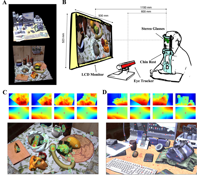
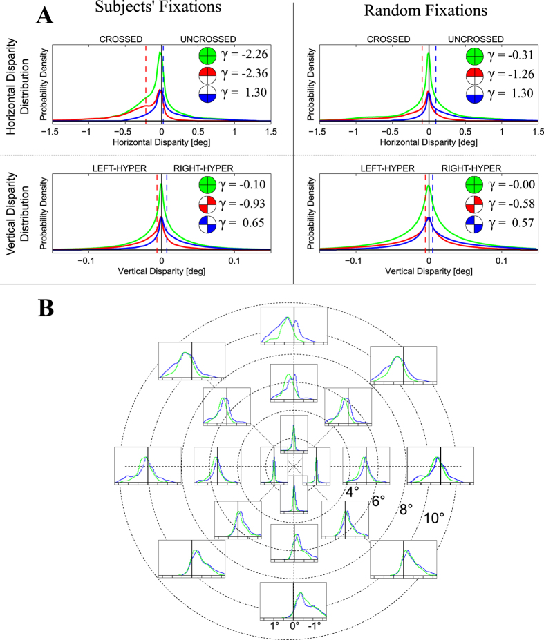
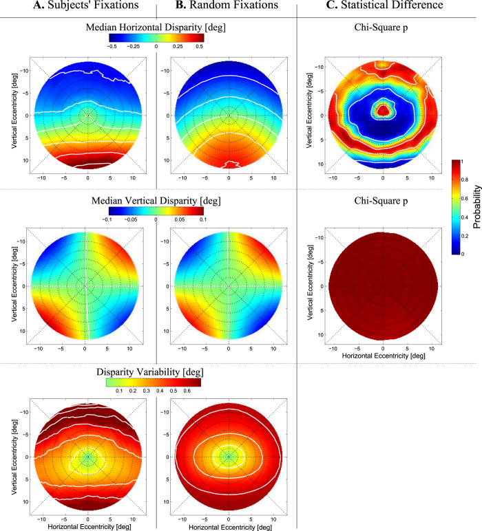
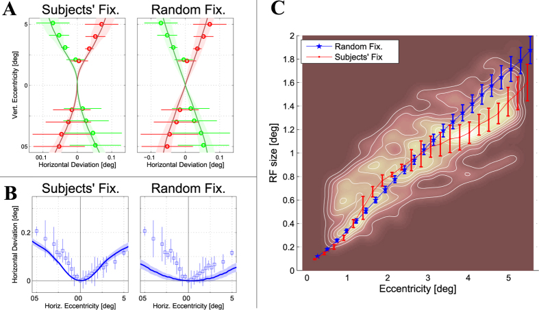
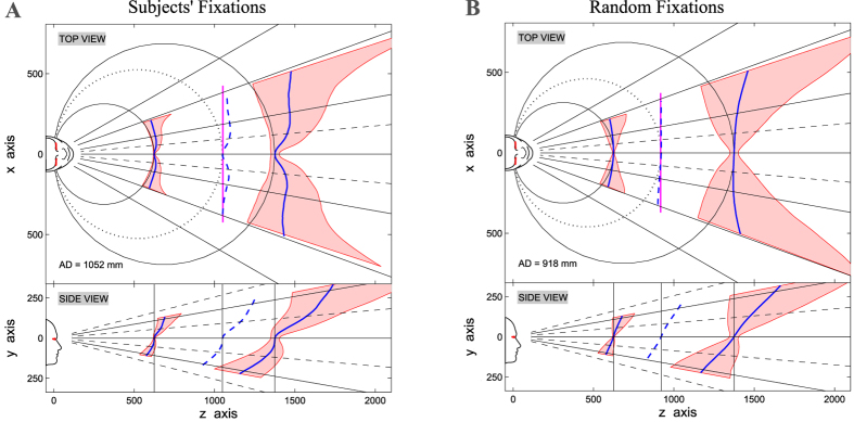
Depth perception in near viewing strongly relies on the interpretation of binocular retinal disparity to obtain stereopsis. Statistical regularities of retinal disparities have been claimed to greatly impact on the neural mechanisms that underlie binocular vision, both to facilitate perceptual decisions and to reduce computational load. In this paper, we designed a novel and unconventional approach in order to assess the role of fixation strategy in conditioning the statistics of retinal disparity. We integrated accurate realistic three-dimensional models of natural scenes with binocular eye movement recording, to obtain accurate ground-truth statistics of retinal disparity experienced by a subject in near viewing. Our results evidence how the organization of human binocular visual system is finely adapted to the disparity statistics characterizing actual fixations, thus revealing a novel role of the active fixation strategy over the binocular visual functionality. This suggests an ecological explanation for the intrinsic preference of stereopsis for a close central object surrounded by a far background, as an early binocular aspect of the figure-ground segregation process.


Conflict of interest statement

The authors declare no competing financial interests.

Figures

Figure 1
Figure 1
(A) Three-dimensional environments. Sketch representing the two naturalistic virtual scenes used: an office desk (top) and a kitchen table (bottom). (B) Stereoscopic experimental setup. Representation of the setup for computing binocular fixations on subjects. (C) Fixation density map. Heatmap (contour lines) and fixation points (red dots) superimposed to the cyclopean image for a subject exploring a binocular image (bottom). and horizontal and vertical disparity patterns for selected fixation points (top). (D) Random fixations. Random fixation points (red dots) (bottom), and selected horizontal and vertical disparity patterns (top).
Figure 2
Figure 2
(A) Disparity distribution on different quadrants. Horizontal (top) and vertical (bottom) disparity distribution for subjects’ (left) and random fixations (right), computed over the whole field of view (green). For horizontal disparity, the distribution has been recomputed separating the upper (red) by the lower (blue) hemifields. For vertical disparity the distribution has been recomputed separating the top-right and bottom-left quadrants (red) by the upper-left and lower-right (blue). According to our notation, crossed horizontal disparities and right-hyper vertical disparity are positive, while uncrossed horizontal disparities and left-hyper vertical disparities are negative. The vertical dashed lines represent the median of the considered distributions. The insets represent the separation, together with the skewness γ of the corresponding distribution. (B) Topographic representation of the disparity distribution. Horizontal disparity distribution computed at different image locations (i.e. pixel) for random fixations (blue) and subjects’ actual fixations (green): three different eccentricities (1°, 5°, 10°) and eight different orientations.
Figure 3
Figure 3
(A) Disparity patterns for subjects’ fixations. Retinotopic patterns of disparity distributions derived from subjects’ fixations: median horizontal (top) and vertical (middle), and standard deviation (bottom). The white lines represent iso-contour levels at steps of 0.1°. (B) Disparity patterns for random fixations. Same representation as in panel A, but derived from random fixations. According to our notation, crossed horizontal disparities and right-hyper vertical disparity are positive (red), while uncrossed horizontal disparities and left-hyper vertical disparities are negative (blue). (C) Distance between the two distributions. χ2 probability (p-value) of the Mahalanobis distance between the random and the subjects’ fixations distributions, for horizontal (left) and vertical (right) disparities.
Figure 4
Figure 4
(A) Helmholtz shear deviation. Helmholtz shear deviation inferred by the obtained disparity distribution (solid lines) for the subjects’ (left) and random (right) fixations. The solid lines and the colored regions represent the mean and standard deviation computed among the four subjects (left), while for random fixations (right)they represent the mean and the 95% confidence interval (1000 bootstraps). The dots and errorbars report the Helmholtz shear deviation (mean and standard deviation) measured in psychophysical experiments from ref. (28 subjects) and ref. (20 subjects). (B) Hering-Hillebrand deviation. Hering-Hillebrand deviation inferred by the obtained disparity distribution (solid lines) for the subjects’ (left) and random (right) fixations. The squares and errorbars report the Hering-Hillebrand deviation (mean and standard deviation) measured from psychophysical measurements of retinal corresponding points from ref. (3 subjects). (C) Receptive field size versus retinal eccentricity. Mean and standard deviation (solid lines and errorbars) of receptive field size (y-axis) with respect to retinal eccentricity (x-axis), inferred by the obtained disparity distribution, among the different subjects (red) and random bootstraps (blue). The background contour map reports the population receptive field size measured from ref. (12 subjects).
Figure 5
Figure 5
(A) Disparity-based prediction of 3D horopter and Panum’s fusional area for subjects’ fixations. The disparity statistics obtained by subjects’ fixations were used to infer a plausible shape for the vertical and horizontal horopter, as well as for Panum’s areas (top and side view). The horopter (blue lines) is computed as the surfaces that projects with minimum error to the pairs of predicted corresponding points. Similarly, the near and far limits of Panum’s area (red regions) were computed as the 15th and the 85th percentile of the disparity distribution. The observer is fixating straight-ahead, with a vergence angle of 7° (≈500 mm). The top view shows for reference the isovergence (Vieth-Müller) circles for the considered vergence angles, and the isoversion lines (gray lines). The dashed blue lines represent the predicted horopters computed at abathic distances, i.e. 3.25° vergence angle (≈1052 mm), with its the best linear fit (magenta line) and the related Vieth-Müller circle (gray dotted line). For a representation of the corresponding 3D horopter, see Supplementary Fig. S3. (B) Disparity-based prediction of 3D horopter and Panum’s fusional area for random fixations. The disparity statistics resulting from random fixations where used to infer the vertical and horizontal horopters and Panum’s area, as in Panel A. The abathic distance corresponds to 3.7° vergence angle (≈918 mm).
Figure 6
Figure 6. Geometrical sketch of thetoe-in technique.
The left and right camera frames: (XL, YL, ZL) and (XR, YR, ZR). The left and right image planes: (xL, yL) and (xR, yR). The camera optical axes are directed towards the fixation point F. b represents the baseline, i.e. the interocular distance. (H, V, T)L/R represents the azimuth, elevation and torsion angles for the left and right cameras, respectively.

Comment on

References

    1. Ohzawa I., Freeman R. D. & DeAngelis G. C. Stereoscopic depth discrimination in the visual cortex: Neurons ideally suited as disparity detectors. Science 249, 1037–1041 (1990). - PubMed
    1. Qian N. Computing stereo disparity and motion with known binocular cell properties. Neural Computation 6(3), 390–404 (1994).
    1. Gilaie-Dotan S., Ullman S., Kushnir T. & Malach R. Shape-selective stereo processing in human object-related visual areas. Human brain mapping 15, 67–79 (2002). - PMC - PubMed
    1. Cottereau B. R., McKee S. P., Ales J. M. & Norcia A. M. Disparity-tuned population responses from human visual cortex. The Journal of Neuroscience 31, 954–965 (2011). - PMC - PubMed
    1. Yang Z. & Purves D. A statistical explanation of visual space. Nature neuroscience 6, 632–640 (2003). - PubMed

Publication types

LinkOut - more resources