

The Dual Nature of Early-Life Experience on Somatosensory Processing in the Human Infant Brain
- PMID: 28318973
- PMCID: PMC5388002
- DOI: 10.1016/j.cub.2017.02.036
The Dual Nature of Early-Life Experience on Somatosensory Processing in the Human Infant Brain
Abstract
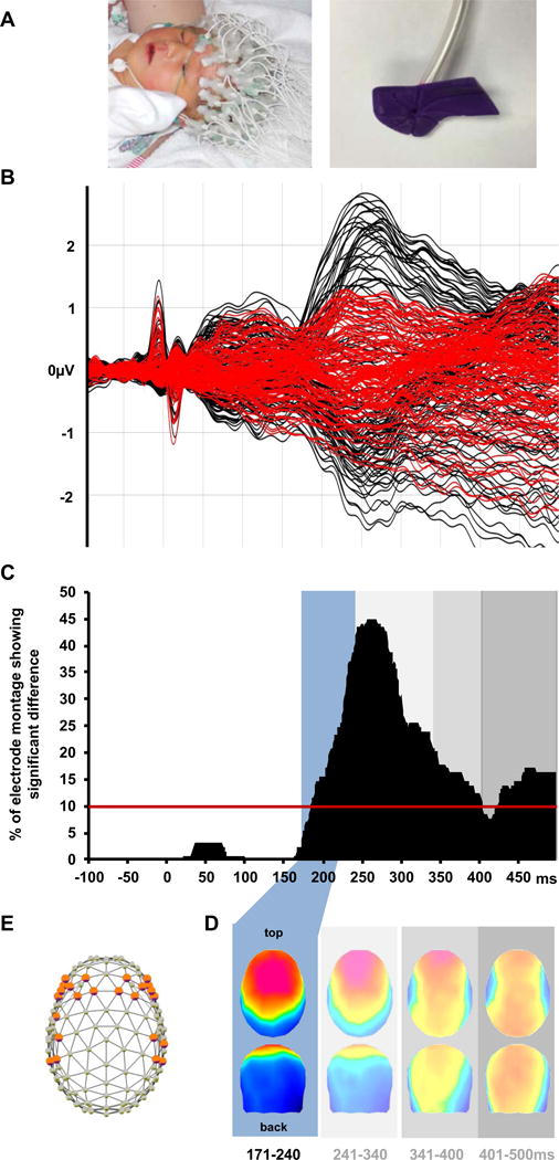
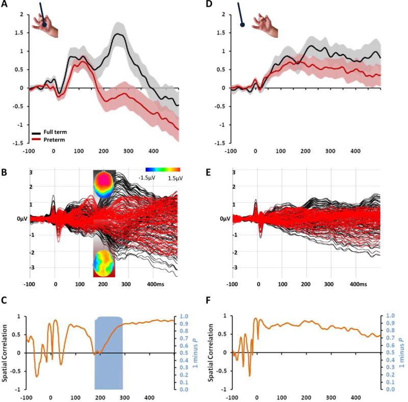
Every year, 15 million preterm infants are born, and most spend their first weeks in neonatal intensive care units (NICUs) [1]. Although essential for the support and survival of these infants, NICU sensory environments are dramatically different from those in which full-term infants mature and thus likely impact the development of functional brain organization [2]. Yet the integrity of sensory systems determines effective perception and behavior [3, 4]. In neonates, touch is a cornerstone of interpersonal interactions and sensory-cognitive development [5-7]. NICU treatments used to improve neurodevelopmental outcomes rely heavily on touch [8]. However, we understand little of how brain maturation at birth (i.e., prematurity) and quality of early-life experiences (e.g., supportive versus painful touch) interact to shape the development of the somatosensory system [9]. Here, we identified the spatial, temporal, and amplitude characteristics of cortical responses to light touch that differentiate them from sham stimuli in full-term infants. We then utilized this data-driven analytical framework to show that the degree of prematurity at birth determines the extent to which brain responses to light touch (but not sham) are attenuated at the time of discharge from the hospital. Building on these results, we showed that, when controlling for prematurity and analgesics, supportive experiences (e.g., breastfeeding, skin-to-skin care) are associated with stronger brain responses, whereas painful experiences (e.g., skin punctures, tube insertions) are associated with reduced brain responses to the same touch stimuli. Our results shed crucial insights into the mechanisms through which common early perinatal experiences may shape the somatosensory scaffolding of later perceptual, cognitive, and social development.
Keywords: development; infant; pain; parent; preterm; sensory; tactile; touch.
Copyright © 2017 Elsevier Ltd. All rights reserved.
Conflict of interest statement
We have no conflicts of interest to report.
Figures
References
-
- Lawn JE, Davidge R, Paul V, Xylander Von C. Born Too Soon: The Global Action Report on Preterm Birth. World Health Organization; 2012. http://www.who.int/pmnch/media/news/2012/20120522_joylawn_presentation.p....
-
- Pineda RG, Neil J, Dierker D, Smyser CD, Wallendorf M, Kidokoro H, Reynolds LC, Walker S, Rogers C, Mathur AM, et al. Alterations in brain structure and neurodevelopmental outcome in preterm infants hospitalized in different neonatal intensive care unit environments. J Pediatr. 2014;164:52–60.e52. - PMC - PubMed
-
- Hrbek A, Karlberg P, Olsson T. Development of visual and somatosensory evoked responses in pre-term newborn infants. Electroencephalography and Clinical Neurophysiology. 1973;34:225–32. 1973. - PubMed
-
- Vanhatalo S, Lauronen L. Neonatal SEP – back to bedside with basic science. Semin Fetal Neonatal Med. 2006;11:464–470. - PubMed
-
- Spittle AJ, Walsh J, Olsen JE, McInnes E, Eeles AL, Brown NC, Anderson PJ, Doyle LW, Cheong JLY. Neurobehaviour and neurological development in the first month after birth for infants born between 32–42 weeks’ gestation. Early Hum Dev. 2016;96:7–14. - PubMed
MeSH terms
Grants and funding
LinkOut - more resources
Full Text Sources
Other Literature Sources
Medical
