. 2017 Apr 3;27(7):1068-1073.
doi: 10.1016/j.cub.2017.01.025. Epub 2017 Mar 16.

Assembly of CNS Nodes of Ranvier in Myelinated Nerves Is Promoted by the Axon Cytoskeleton

Affiliations

Assembly of CNS Nodes of Ranvier in Myelinated Nerves Is Promoted by the Axon Cytoskeleton

Veronica Brivio et al. Curr Biol. .

Abstract

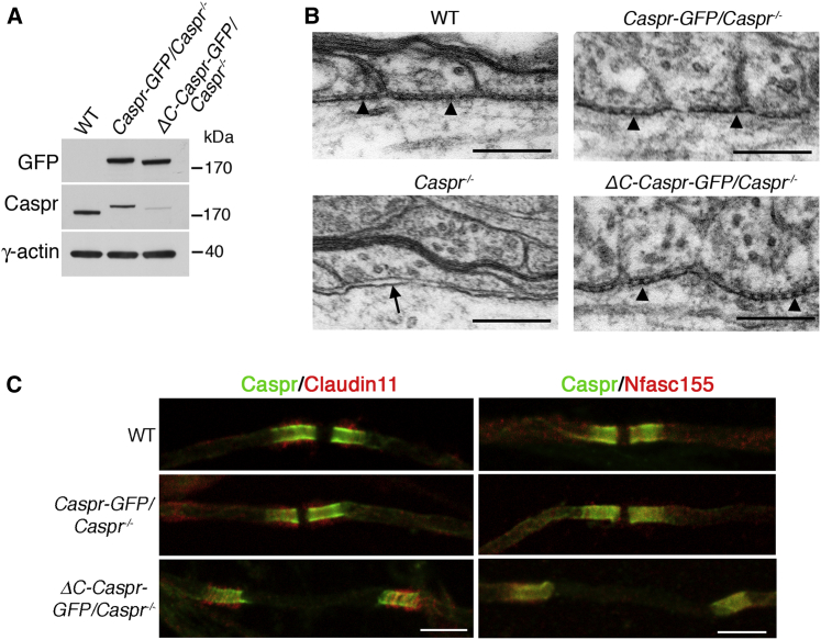
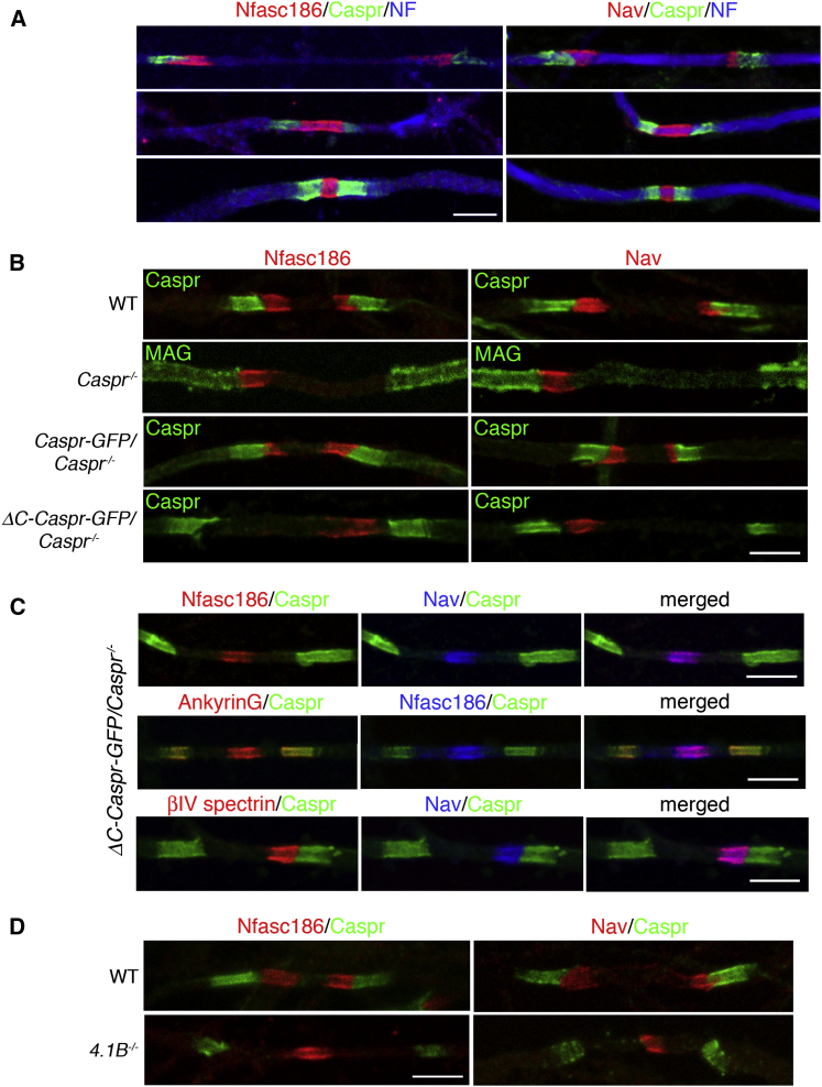
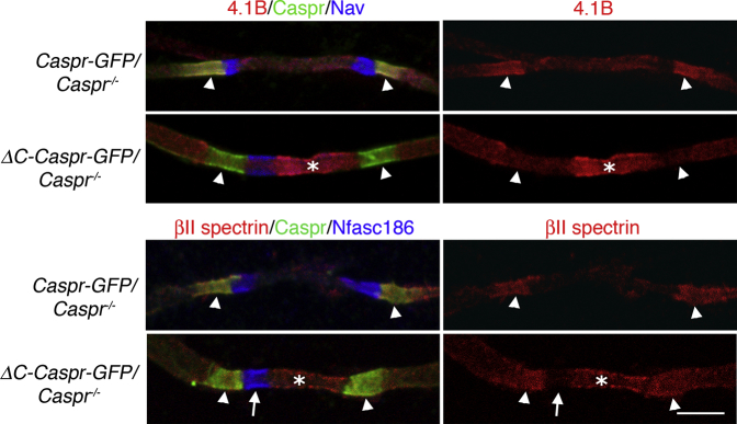
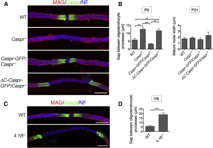
Nodes of Ranvier in the axons of myelinated neurons are exemplars of the specialized cell surface domains typical of polarized cells. They are rich in voltage-gated sodium channels (Nav) and thus underpin rapid nerve impulse conduction in the vertebrate nervous system [1]. Although nodal proteins cluster in response to myelination, how myelin-forming glia influence nodal assembly is poorly understood. An axoglial adhesion complex comprising glial Neurofascin155 and axonal Caspr/Contactin flanks mature nodes [2]. We have shown that assembly of this adhesion complex at the extremities of migrating oligodendroglial processes promotes process convergence along the axon during central nervous system (CNS) node assembly [3]. Here we show that anchorage of this axoglial complex to the axon cytoskeleton is essential for efficient CNS node formation. When anchorage is disrupted, both the adaptor Protein 4.1B and the cytoskeleton protein βII spectrin are mislocalized in the axon, and assembly of the node of Ranvier is significantly delayed. Nodal proteins and migrating oligodendroglial processes are no longer juxtaposed, and single detached nodal complexes replace the symmetrical heminodes found in both the CNS and peripheral nervous system (PNS) during development. We propose that axoglial adhesion complexes contribute to the formation of an interface between cytoskeletal elements enriched in Protein 4.1B and βII spectrin and those enriched in nodal ankyrinG and βIV spectrin. This clusters nascent nodal complexes at heminodes and promotes their timely coalescence to form the mature node of Ranvier. These data demonstrate a role for the axon cytoskeleton in the assembly of a critical neuronal domain, the node of Ranvier.

Keywords: CNS; myelination; node of Ranvier; paranodal axoglial junction.


Figures

Figure 1
Figure 1
Extracellular Axoglial Adhesion in Caspr Null Mice Expressing Caspr without Its C Terminus (A) Western blotting of spinal cord lysates on a 4%–12% NuPAGE gel showed that expression levels of Caspr-GFP and ΔC-Caspr-GFP were similar in Caspr null mice. Samples (20 μg) were blotted sequentially with mouse anti-GFP followed by rabbit anti-Caspr, with γ-actin blotted as a loading control in each lane. As expected, Caspr-GFP appears larger than WT Caspr and ΔC-Caspr-GFP is smaller than Caspr-GFP. (B) Electron microscopy of paranodes from the ventral funiculus of the spinal cord of WT, Caspr-GFP/Caspr−/−, and ΔC-Caspr-GFP/Caspr−/− mice at P41 showed the extracellular adhesion complex at the axoglial interface as electron-dense septate junctions. In Caspr−/− mice septate junctions were absent. The scale bars represent 1 μm. (C) Immunofluorescence at P6 showed that Caspr, Caspr-GFP, and ΔC-Caspr-GFP colocalized with both the oligodendroglial paranodal loop marker Claudin 11 and the adhesion complex protein Nfasc155. The scale bars represent 5 μm.
Figure 2
Figure 2
Mislocalization of Nodal Complexes in Caspr Null and ΔC-Caspr-GFP/Caspr−/− Mice (A) Although most nodes are mature in WT nerves at P6 (see Figure 4), earlier stages of heminode fusion can be detected by immunofluorescence for either Nfasc186 or Nav (both nodal proteins) with Caspr and Neurofilament (NF). (B) Immunofluorescence at P6 showed that complete disruption of the axoglial complex (Caspr−/−) or removal of the Protein 4.1B binding site in Caspr (ΔC-Caspr-GFP/Caspr−/−) abolished symmetrical heminode formation and resulted in a single nodal complex. Heminode formation was rescued with transgenic Caspr (Caspr-GFP/Caspr−/−). (C) Immunostaining at P6 showed that the mislocalized nodal complex in ΔC-Caspr-GFP/Caspr−/− mice contained Nav, Nfasc186, βIV spectrin, and ankyrinG. (D) Immunofluorescence at P6 for Nfasc186 and Nav in the absence of Protein 4.1B (4.1B−/−) showed that the nodal complex was mislocalized. The scale bars represent 5 μm. See also Figure S1.
Figure 3
Figure 3
Mislocalization of Axonal Protein 4.1B and βII Spectrin in ΔC-Caspr-GFP/Caspr−/− Mice Immunostaining at P6 showed depletion of Protein 4.1B at the axoglial junction in ΔC-Caspr-GFP/Caspr−/− mice (arrowheads) and concomitant invasion of the protein into the axon between converging processes (asterisk). Although βII spectrin persisted at the axoglial junction (arrowheads) in ΔC-Caspr-GFP/Caspr−/− mice, it was also mislocalized between converging preocesses (asterisk). Nevertheless, it was still largely excluded from the nodal complex (arrow), as observed in control Caspr-GFP/Caspr−/− mice. The scale bar represents 5 μm.
Figure 4
Figure 4
Interaction of the Intracellular Domain of Caspr with Axonal Protein 4.1B Promotes Oligodendroglial Process Migration (A) Immunofluorescence at P6 for Myelin-Associated Glycoprotein (MAG), the axonal marker Neurofilament-H (NF), Caspr-GFP, and ΔC-Caspr-GFP suggested that the migration of oligodendroyte processes was delayed in Caspr−/− and ΔC-Caspr-GFP/Caspr−/− mice. (B) Quantitation of the gaps between the tips of migrating processes at P6 and P21. Caspr-GFP rescued the delay in process migration observed in Caspr−/− mice at P6 whereas ΔC-Caspr-GFP did not. However, by P21 there was no significant difference in the width of the nodal gap among the four genotypes. Values are means ± SEM (n = 3 mice per genotype, a minimum of 50 gaps between converging pairs of processes were measured per mouse; ∗∗∗p ≤ 0.001 and ∗∗p ≤ 0.01, Tukey’s multiple comparison test). (C and D) Immunofluorescence (C) and quantitative analysis (D) of Protein 4.1B null (4.1B−/−) spinal cord fibers at P6 (as in A and B) showed that the convergence of oligodendroglial processes was also significantly delayed compared with WT fibers. Values are means ± SEM (n = 3 mice per genotype, a minimum of 50 gaps between converging pairs of processes were measured per mouse; ∗∗∗p ≤ 0.001, unpaired Student’s t test). The scale bars represent 5 μm.

References

    1. Huxley A.F., Stämpfli R. Evidence for saltatory conduction in peripheral myelinated nerve fibres. J. Physiol. 1949;108:315–339. - PubMed
    1. Sherman D.L., Tait S., Melrose S., Johnson R., Zonta B., Court F.A., Macklin W.B., Meek S., Smith A.J., Cottrell D.F., Brophy P.J. Neurofascins are required to establish axonal domains for saltatory conduction. Neuron. 2005;48:737–742. - PubMed
    1. Zonta B., Tait S., Melrose S., Anderson H., Harroch S., Higginson J., Sherman D.L., Brophy P.J. Glial and neuronal isoforms of Neurofascin have distinct roles in the assembly of nodes of Ranvier in the central nervous system. J. Cell Biol. 2008;181:1169–1177. - PMC - PubMed
    1. Bhat M.A., Rios J.C., Lu Y., Garcia-Fresco G.P., Ching W., St Martin M., Li J., Einheber S., Chesler M., Rosenbluth J. Axon-glia interactions and the domain organization of myelinated axons requires neurexin IV/Caspr/Paranodin. Neuron. 2001;30:369–383. - PubMed
    1. Boyle M.E., Berglund E.O., Murai K.K., Weber L., Peles E., Ranscht B. Contactin orchestrates assembly of the septate-like junctions at the paranode in myelinated peripheral nerve. Neuron. 2001;30:385–397. - PubMed

LinkOut - more resources