. 2017 Jul 20;36(29):4089-4099.
doi: 10.1038/onc.2017.11. Epub 2017 Mar 20.

Iron addiction: a novel therapeutic target in ovarian cancer

Affiliations

Iron addiction: a novel therapeutic target in ovarian cancer

D Basuli et al. Oncogene. .

Abstract

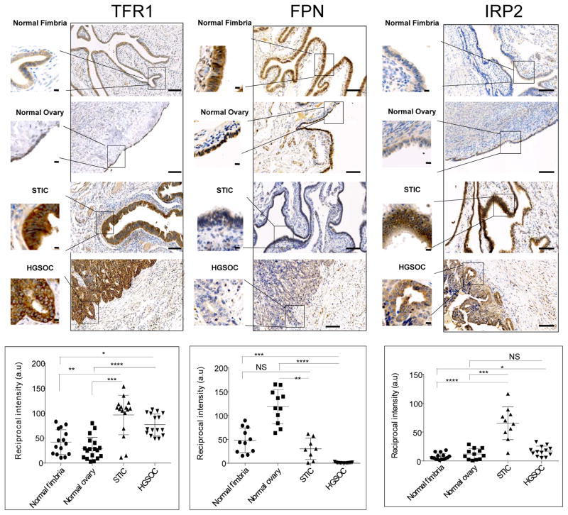
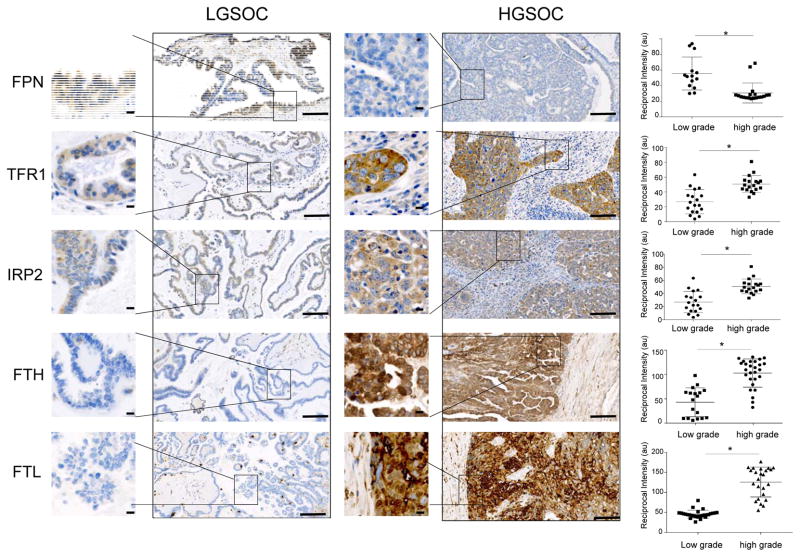
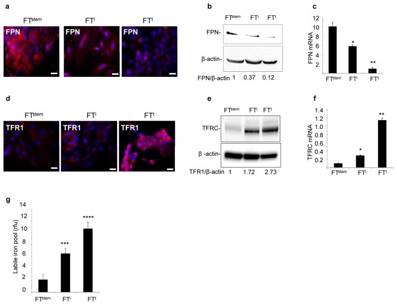
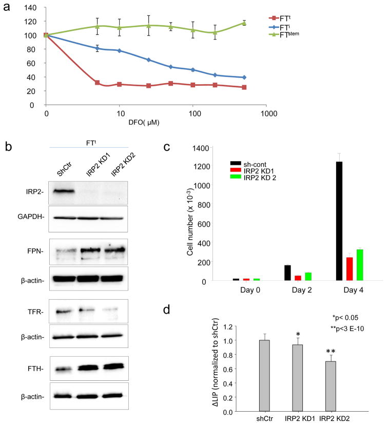
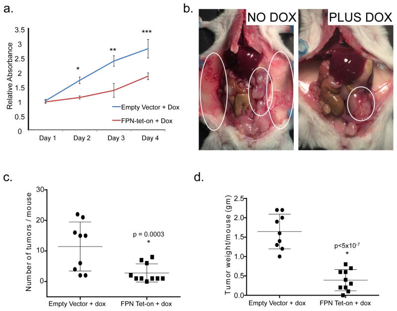
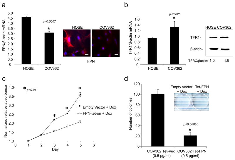
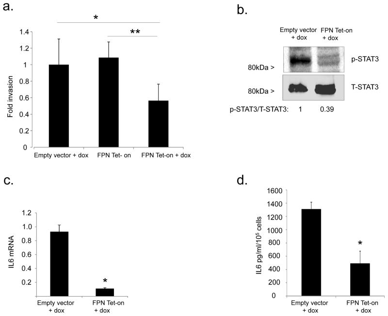
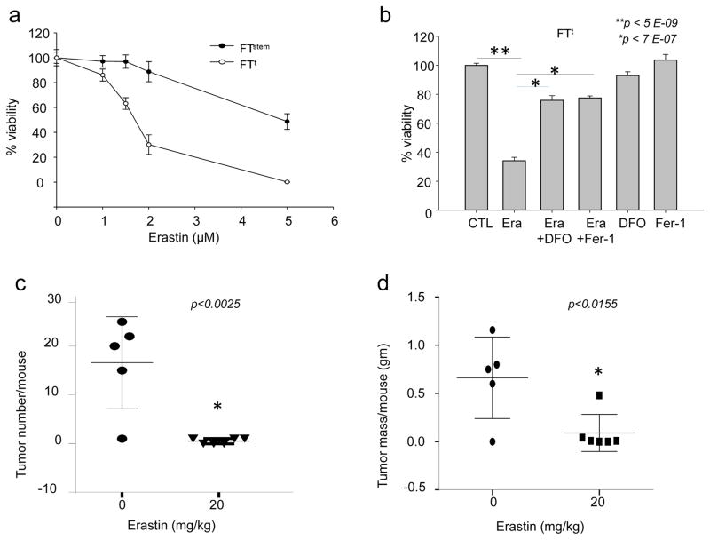
Ovarian cancer is a lethal malignancy that has not seen a major therapeutic advance in over 30 years. We demonstrate that ovarian cancer exhibits a targetable alteration in iron metabolism. Ferroportin (FPN), the iron efflux pump, is decreased, and transferrin receptor (TFR1), the iron importer, is increased in tumor tissue from patients with high grade but not low grade serous ovarian cancer. A similar profile of decreased FPN and increased TFR1 is observed in a genetic model of ovarian cancer tumor-initiating cells (TICs). The net result of these changes is an accumulation of excess intracellular iron and an augmented dependence on iron for proliferation. A forced reduction in intracellular iron reduces the proliferation of ovarian cancer TICs in vitro, and inhibits both tumor growth and intraperitoneal dissemination of tumor cells in vivo. Mechanistic studies demonstrate that iron increases metastatic spread by facilitating invasion through expression of matrix metalloproteases and synthesis of interleukin 6 (IL-6). We show that the iron dependence of ovarian cancer TICs renders them exquisitely sensitive in vivo to agents that induce iron-dependent cell death (ferroptosis) as well as iron chelators, and thus creates a metabolic vulnerability that can be exploited therapeutically.


Conflict of interest statement

Conflict of interest statement. The authors declare no conflict of interest.

Figures

Fig. 1
Fig. 1. Proteins that control intracellular iron are altered high grade serous ovarian cancer (HGSOC)
Representative images of immunohistochemical staining of normal fimbria, normal ovary surface epithelia, serous tubal intra-epithelial carcinoma (STIC) and HGSOC stained with antibodies against transferrin receptor 1 (TFR1), ferroportin (FPN) and iron regulatory protein 2 (IRP2). Dot plots represent quantification of staining of tissues collected from 8 patients with HGSOC and 5 with STIC compared to 8 subjects with normal fimbria and 6 individuals with normal ovarian surface epithelium. Images of three to four random fields per slide were quantified. Differences in TRFC (p<0.0001), FPN (p<0.0001 and IRP2 (p<0.01) were statistically significant (one way ANOVA). *p<0.001, **p<5x10⁻⁵, *** p<5x10⁻⁶, ****p<5x10⁻⁷, one tailed t test for individual comparisons. Scale bar 1 mm; inset scale bar 10 μm.
Fig. 2
Fig. 2. Proteins that control intracellular iron differ in high grade and low grade serous ovarian cancer
Representative images of immunohistochemical staining of tumor tissues from patients with low grade serous ovarian cancer (LGSOC) and high grade serous ovarian cancer (HGSOC). Proteins stained are ferroportin (FPN), transferrin receptor (TFR1), Iron Regulatory Protein 2 (IRP2), ferritin heavy chain (FTH) and ferritin light chain (FTL). Dot plots show quantification of staining of tissues from 5 patients with LGSOC and 8 patients with HGSOC (3 to 4 random fields from each patient tissue slide). *p<0.002. Scale bar 1 mm; inset scale bar 10 μm.
Fig. 3
Fig. 3. Iron metabolism is modified in a genetic model of ovarian tumor initiating cells
(a,d) Immunofluorescent staining of ferroportin (FPN) and transferrin receptor (TFR1) in normal fallopian tube stem cells (FTstem), immortalized fallopian tube stem cells (FTi) and transformed fallopian tube stem cells (FTt). FPN and TFR1 in red; nuclei in blue. Scale bar 20 μm. (b,e)Cropped images of western blots and quantification of FPN and TFR1 with β-actin as a loading control. (c,f) qRT-PCR of FPN mRNA and TFR1 mRNA in FTstem, FTi and FTt cells. (n) Labile iron pool (LIP) per 1000 cells. *p<0.02, **p<0.0.0007, ***p<5x10⁻⁸; ****p<5x10⁻¹¹.
Fig. 4
Fig. 4. Tumor-initiating cells exhibit increased iron dependence
(a) Cells were treated for 72 hrs with the indicated concentrations of deferoxamine (DFO) and viability was assessed using an MTS assay. (b) Cropped images of western blots of IRP2, transferrin receptor, (TFR1) ferritin heavy chain (FTH), and ferroportin in FTt cells with knockdown of IRP2 (IRP2 KD1, IRP2KD2) or control shRNA (shCtr). (c) Cell proliferation as assessed by trypan-blue exclusion in FTt cells treated with control shRNA or IRP2 knockdown vectors. d) Labile iron pool in cells with knockdown of IRP2 or control shRNA
Fig. 5
Fig. 5. Increased iron efflux decreases tumorigenicity and invasion of ovarian cancer tumor-initiating cells
(a) Cell viability of FTt cells as assayed by MTS assay at indicated time points with and without ferroportin overexpression. Blue line represents control (empty vector with doxycycline), and red line represents FPN-tet-on FTt cells (ferroportin tet on with doxycycline). *p<0.0002, one tail t test. (b) Representative images of mice inoculated IP with FPN-tet-on FTt cells and left untreated (left) or treated with doxycycline (right) for four weeks to induce expression of ferroportin. Untreated mice have a greater tumor burden and wider area of metastasis (circled in white). (c) Quantification of tumor number/mouse and (d) tumor mass/mouse following implantation of tumor cells containing empty vector (n=9) or FPN tet-on (n=10) following 4 weeks of doxycycline treatment.
Fig. 6
Fig. 6. Increased iron efflux reduces proliferation of COV362 ovarian cancer cells
(a) q-RTPCR of FPN (normalized to βactin) and immunofluorescence staining of FPN in COV362 and HOSE cells: FPN in red; nuclei in blue. Scale bar 20 μm. (b)q-RTPCR of TFR1/βactin in COV362 ovarian cancer cells and HOSE cells; (c) FPN was induced at time 0 by the addition of doxycycline and cell viability assessed at the indicated timepoints by MTS assay; (e) Colony formation of COV362cells with and without ferroportin overexpression was analyzed by crystal violet staining. Colonies from three replicate wells were counted and quantified.
Fig. 7
Fig. 7. Ferroportin modulates invasion of FTt cells through STAT3 and IL6
(a) In vitro invasion was measured in a simplified Boyden chamber consisting of two chambers separated by an 8 micron porous filter coated with or without basement membrane extract (BME). Invasion was normalized to migration of the cells in the inserts without the BME coating. *p=0.002; **p=5x10⁻⁵. (b) Western blot analysis of p-STAT3 and total STAT-3 in FTt cells transduced with empty vector (EV) or FPN-Tet-on following 48 hrs exposure to doxycycline. Band intensities were quantified using ImageJ. (c) IL6 mRNA was assessed using qRT-PCR; (d) IL6 was measured in culture supernates following 48 hrs’ exposure to doxycycline. *p≤0.0002.
Fig. 8
Fig. 8. Erastin induces ferroptosis in tumor-initiating cells and reduces tumor number and mass in vivo
(a) 3000 FTstem and FTt cells were treated with indicated doses of erastin and cell viability assessed 48 hours later. (b) FTt cells were treated for 48 hrs with 2 μM of erastin alone, the combination of 2uM erastin plus 2 μM ferrostatin-1, the combination of 2uM erastin plus 50 uM DFO, ferrostatin alone, or DFO alone and cell viability assessed. (c,d) Mice were injected intraperitoneally with FTt cells and treated for 18 days with either vehicle or 20 mg/kg erastin. The number of tumors/mouse and total tumor mass/mouse were measured in the vehicle-treated group (n=5) and in the group treated with erastin (n=6).

References

    1. A snapshot of ovarian cancer. [cited 2015 December 21 2015]. Available from: http://www.cancer.gov/research/progress/snapshots/ovarian.
    1. Vang R, Shih Ie M, Kurman RJ. Ovarian low-grade and high-grade serous carcinoma: pathogenesis, clinicopathologic and molecular biologic features, and diagnostic problems. Adv Anat Pathol. 2009 Sep;16(5):267–82. Epub 2009/08/25. eng. - PMC - PubMed
    1. Leong HS, Galletta L, Etemadmoghadam D, George J, Kobel M, Ramus SJ, et al. Efficient molecular subtype classification of high-grade serous ovarian cancer. J Pathol. 2015 Jul;236(3):272–7. Epub 2015/03/27. eng. - PubMed
    1. Tothill RW, Tinker AV, George J, Brown R, Fox SB, Lade S, et al. Novel molecular subtypes of serous and endometrioid ovarian cancer linked to clinical outcome. Clin Cancer Res. 2008 Aug 15;14(16):5198–208. Epub 2008/08/14. eng. - PubMed
    1. Lengyel E. Ovarian cancer development and metastasis. Am J Pathol. 2010 Sep;177(3):1053–64. Epub 2010/07/24. eng. - PMC - PubMed

Publication types