. 2017 Mar 20;12(3):e0173726.
doi: 10.1371/journal.pone.0173726. eCollection 2017.

Subchondral bone histology and grading in osteoarthritis

Affiliations

Subchondral bone histology and grading in osteoarthritis

Olli-Matti Aho et al. PLoS One. .

Abstract

Objective: Osteoarthritis (OA) has often regarded as a disease of articular cartilage only. New evidence has shifted the paradigm towards a system biology approach, where also the surrounding tissue, especially bone is studied more vigorously. However, the histological features of subchondral bone are only poorly characterized in current histological grading scales of OA. The aim of this study is to specifically characterize histological changes occurring in subchondral bone at different stages of OA and propose a simple grading system for them.

Design: 20 patients undergoing total knee replacement surgery were randomly selected for the study and series of osteochondral samples were harvested from the tibial plateaus for histological analysis. Cartilage degeneration was assessed using the standardized OARSI grading system, while a novel four-stage grading system was developed to illustrate the changes in subchondral bone. Subchondral bone histology was further quantitatively analyzed by measuring the thickness of uncalcified and calcified cartilage as well as subchondral bone plate. Furthermore, internal structure of calcified cartilage-bone interface was characterized utilizing local binary patterns (LBP) based method.

Results: The histological appearance of subchondral bone changed drastically in correlation with the OARSI grading of cartilage degeneration. As the cartilage layer thickness decreases the subchondral plate thickness and disorientation, as measured with LBP, increases. Calcified cartilage thickness was highest in samples with moderate OA.

Conclusion: The proposed grading system for subchondral bone has significant relationship with the corresponding OARSI grading for cartilage. Our results suggest that subchondral bone remodeling is a fundamental factor already in early stages of cartilage degeneration.


Conflict of interest statement

Competing Interests: The authors have declared that no competing interests exist.

Figures

Fig 1
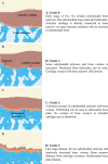
Fig 1. Subchondral bone changes and grades.
(A) Grade 0 Early stage of OA. No evident subchondral bone sclerosis, thin subchondral bone plate and trabeculae. Articular cartilage is directly connected to bone marrow via open fenestrae (marked with an asterisk) in subchondral bone. (B) Grade 1. Some subchondral sclerosis and bone volume is increased. Thickened bone trabeculae can be seen. Cartilage contact with bone marrow still persists. (C) Grade 2. A distinct increase in subchondral sclerosis and bone volume. Fibrillation can be seen in subcondral bone plate. No contact of bone marrow to articular cartilage can be identified. (D) Grade 3. Late stage disease. Severe subchondral sclerosis and massively increased bone volume. Bone marrow distance from cartilage increases. Subchondral bone plate flattens.
Fig 2
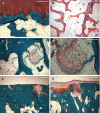
Fig 2. Safranin O (A-D) and Masson’s trichrome stained histological samples of subchondral bone grades.
Images taken with a light microscope using digital camera. White triangle marks articular cartilage, white cross shows calcified cartilage. (A and E) Black asterisks marks fenestrae in subchondral bone plate connecting the articular cartilage to bone marrow in grade 0 and (B and F) grade 1. (C and G) Fibrillation on subchondral bone plate can be seen in grade 2. (D and H) Distinctive sclerosis and loss of articular cartilage mark late-stage OA in grade 3. Scale bar 200 μm.
Fig 3
Fig 3. LBP analysis of the interface bone-cartilage for a sample with early OA.
On left an image of a sample with light degeneration (OARSI: 1.0) and a sample with severe OA on right (OARSI: 4.5). (A) Original image, original magnification x5. (B) Manual segmentation of the interface bone-cartilage and (C) distribution of local angles at the interface from LBP analysis.
Fig 4
Fig 4. Masson’s trichrome (A, C-F) and HE stained (B) histological images of the features of subchondral bone.
(A) Subchondral bone fenestra marked with an asterisk. (B) Subchondral trabeculae showing some intra-trabecular cartilage (triangles), polarized light microscopy. (C-D) Some of the more degenerated samples were seen with cartilage (triangle) among trabecular bone. (E-F) Late-stage degeneration samples revealing degenerated and apoptotic cartilage. Scale bar 200 μm.
Fig 5
Fig 5. Mean OARSI scores of subchondral bone grades and histological image analysis data in correlation to samples bone OA grade.
(A) OARSI score increases by subchondral oa grade. Samples with subchondral grade 0 reached mean OARSI score of 1.46. Subchondral OA grades 1, 2 and 3 had mean OARSI scores of 3.45, 3.99 and 5.19 respectively. Each bar and point represents the mean ± S.E. (B) Mean cartilage thickness. (C) Mean subchondral bone plate thickness. (D) Mean calcified cartilage thickness. Each bar and point represents the mean ± S.E. * p<0.05, *** p<0.001
Fig 6
Fig 6. LBP based homogeneity and entropy of subchondral bone in correlation to bone and cartilage OA grade.
(A) Homogeneity and (B) entropy in samples with subchondral bone grade 0-2. Grade 3 samples had smooth and worn surface and were excluded. (N = 2, 9 and 7 for grades 0, 1 and 2 respectively). Linear regression models between (C) homogeneity, (D) entropy and cartilage OARSI grade, r2(C) = 0.583, r(D) = 0.612. * p<0.05, *** p<0.001.

References

    1. Lories RJ and Luyten FP. The bone-cartilage unit in osteoarthritis. Nat Rev Rheumatol. 2011;7:43–9. 10.1038/nrrheum.2010.197 - DOI - PubMed
    1. Sellam J, Herrero-Beaumont G, Berenbaum F. Osteoarthritis: pathogenesis, clinical aspects and diagnosis. EULAR compendium on rheumatic diseases. Italy; BMJ Publishing Group LTD; 2009;p444–63.
    1. Moskowitz RW. The burden of osteoarthritis: clinical and quality-of-life issues. Am.J.Manag.Care 2009;15:S223–9. - PubMed
    1. Goldring MB, Goldring SR. Articular cartilage and subchondral bone in the pathogenesis of osteoarthritis. Ann N Y Acad Sci. 2010;1192:230–7. 10.1111/j.1749-6632.2009.05240.x - DOI - PubMed
    1. Pastoureau PC, Chomel AC, Bonnet J. Evidence of early subchondral bone changes in the meniscectomized guinea pig. A densitometric study using dual-energy X-ray absorptiometry subregional analysis. OA Cartilage 1999;7:466–73 - PubMed