The impact of high grade glial neoplasms on human cortical electrophysiology
- PMID: 28319187
- PMCID: PMC5358752
- DOI: 10.1371/journal.pone.0173448
The impact of high grade glial neoplasms on human cortical electrophysiology
Abstract
Objective: The brain's functional architecture of interconnected network-related oscillatory patterns in discrete cortical regions has been well established with functional magnetic resonance imaging (fMRI) studies or direct cortical electrophysiology from electrodes placed on the surface of the brain, or electrocorticography (ECoG). These resting state networks exhibit a robust functional architecture that persists through all stages of sleep and under anesthesia. While the stability of these networks provides a fundamental understanding of the organization of the brain, understanding how these regions can be perturbed is also critical in defining the brain's ability to adapt while learning and recovering from injury.
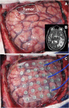
Methods: Patients undergoing an awake craniotomy for resection of a tumor were studied as a unique model of an evolving injury to help define how the cortical physiology and the associated networks were altered by the presence of an invasive brain tumor.
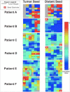
Results: This study demonstrates that there is a distinct pattern of alteration of cortical physiology in the setting of a malignant glioma. These changes lead to a physiologic sequestration and progressive synaptic homogeneity suggesting that a de-learning phenomenon occurs within the tumoral tissue compared to its surroundings.
Significance: These findings provide insight into how the brain accommodates a region of "defunctionalized" cortex. Additionally, these findings may have important implications for emerging techniques in brain mapping using endogenous cortical physiology.
Conflict of interest statement
Figures


References
-
- Biswal B, Yetkin FZ, Haughton VM, Hyde JS. Functional connectivity in the motor cortex of resting human brain using echo-planar MRI. Magn Reson Med. 1995;34(4):537–41. Epub 1995/10/01. - PubMed
-
- Arfanakis K, Cordes D, Haughton VM, Moritz CH, Quigley MA, Meyerand ME. Combining independent component analysis and correlation analysis to probe interregional connectivity in fMRI task activation datasets. Magn Reson Imaging. 2000;18(8):921–30. - PubMed
MeSH terms
Grants and funding
LinkOut - more resources
Full Text Sources
Other Literature Sources
Medical

