. 2017 May:278:8-17.
doi: 10.1016/j.jmr.2017.02.020. Epub 2017 Feb 28.

31P-dephased, 13C-detected REDOR for NMR crystallography at natural isotopic abundance

Affiliations

31P-dephased, 13C-detected REDOR for NMR crystallography at natural isotopic abundance

Alexander I Greenwood et al. J Magn Reson. 2017 May.

Abstract

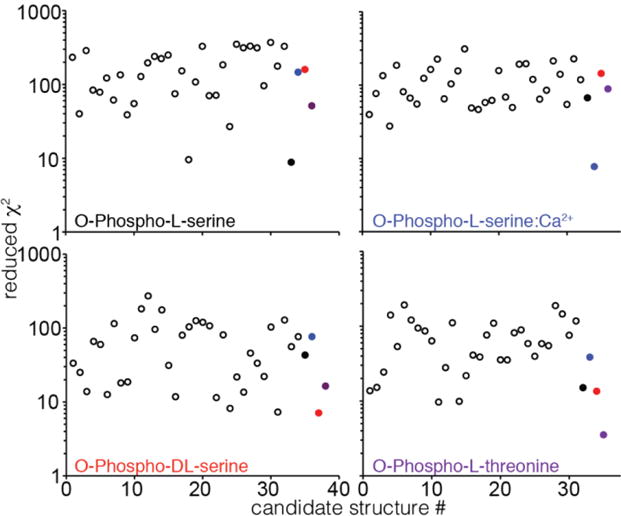
Typically, the process of NMR-based structure determination relies on accurately measuring a large number of internuclear distances to serve as restraints for simulated annealing calculations. In solids, the rotational-echo double-resonance (REDOR) experiment is a widely used approach to determine heteronuclear dipolar couplings corresponding to distances usually in the range of 1.5-8Å. A challenge in the interpretation of REDOR data is the degeneracy of symmetric subunits in an oligomer or equivalent molecules in a crystal lattice, which produce REDOR trajectories that depend explicitly on two or more distances instead of one. This degeneracy cannot be overcome by either spin dilution (for molecules containing 31P, 19F and other highly abundant nuclei) or selective pulses (in the case where there is chemical shift degeneracy). For small, crystalline molecules, such as phosphoserine, we demonstrate that as many as five inter-molecular distances must be considered to model 31P-dephased REDOR data accurately. We report excellent agreement between simulation and experiment once lattice couplings, 31P chemical shift anisotropy, and radio-frequency field inhomogeneity are all taken into account. We also discuss the systematic inaccuracies that may result from approximations that consider only the initial slope of the REDOR trajectory and/or that utilize a two- or three-spin system. Furthermore, we demonstrate the applicability of 31P-dephased REDOR for validation or refinement of candidate crystal structures and show that this approach is especially informative for NMR crystallography of 31P-containing molecules.

Keywords: Magic-angle spinning; Phosphorus; Phosphoserine; Simulation; Solid-state NMR.


Figures

Figure 1
Figure 1. Applications of 31P-dephased REDOR to structural characterization of phosphorus-containing materials
(a–c) Examples of systems that can be characterized using 31P-dephased REDOR: (a) Crystal structure of O-phospho-DL-tyrosine, determined by NMR crystallography. (b) Proteins on the surface of and embedded within a lipid bilayer, composed of lipids with phosphate headgroups. (c) Protein bound to the surface of hydroxyapatite, a mineral composed of inorganic phosphate and calcium. (d) The REDOR pulse sequence, which recouples heteronuclear dipolar couplings with a train of rotor-synchronized π pulses. (e) REDOR trajectories of diluted crystalline N-acetyl-valine (left) and natural-abundance O-phospho-serine (right) with fits to Eq. (1) shown as lines.
Figure 2
Figure 2
Chemical structures of compounds investigated in this study, with α, β, and γ carbons specified.
Figure 3
Figure 3. Measurement of B1 field profiles, 31P CSA, and multi-spin effects
(a) 31P nutation curve of O-phospho-L-serine sample after initial 1H-31P CP. (b) Resultant B1 field profiles for O-phospho-L-serine (black), O-phospho-DL-serine (red), O-phospho-L-serine:Ca2+ (blue) and O-phospho-L-threonine (purple) samples. (c) 31P sideband pattern of O-phospho-L-serine at 4 kHz spinning with vertically-offset fits in red. (d) X-ray structure of the crystal lattice of O-phospho-L-serine, with phosphate groups of six closest 31P nuclei to a given carboxyl carbon emphasized, numbered according to their distance to the carboxyl carbon. (e) Simulated O-phospho-L-serine CO REDOR trajectories including the closest 1, 2, 3, 4, 5, or 6 31P atoms to the CO carbon, as shown in (c). (f) Reduced χ2 between CO REDOR data and simulations as a function of the number of included 31P spins, with RF inhomogeneity and CSA simulated.
Figure 4
Figure 4. Relative contributions of lattice couplings, CSA, and RF inhomogeneity on simulated 31P-dephased REDOR trajectories
(a) Models of increasing complexity used to simulate REDOR trajectories. (b) Comparison of experimental CO REDOR data (symbols) and simulations (solid lines) using different models specified in (a), on various forms of phosphoserine and phosphothreonine. Error bars are standard error on the mean of either 6 or 8 replicates. Reduced χ2 between data and simulation is shown for each trajectory.
Figure 5
Figure 5. Discrimination between different forms of phosphoserine and phosphothreonine by REDOR trajectories
REDOR data (circles) for each carbon of O-phospho-L-serine (black), O-phospho-DL-serine (red), O-phospho-L-serine:Ca2+ (blue) and O-phospho-L-threonine (purple) compared with simulations (solid lines). For the weaker couplings (CO, C α) the REDOR trajectories for each molecule are easily distinguishable from each other and are reproduced well by simulations. Error bars are standard error on the mean of either 6 or 8 replicates. Reduced χ2 between data and simulation is shown for each trajectory.
Figure 6
Figure 6. Discrimination between incorrect candidate structures and published structures by REDOR trajectories
Reduced χ2 between REDOR data and simulation is shown for synthetically generated crystal structures (open circles) and published structures (closed circles, O-phospho-L-serine in black, O-phospho-DL-serine in red, O-phospho-L-serine:Ca2+ in blue, O-phospho-L-threonine in purple). In each case the lowest χ2 corresponds to the published structure.

References

    1. Oldfield E. Quantum chemical studies of protein structure. Philos T R Soc B. 2005;360:1347–1361. - PMC - PubMed
    1. Sun HH, Sanders LK, Oldfield E. Carbon-13 NMR shielding in the twenty common amino acids: Comparisons with experimental results in proteins. J Am Chem Soc. 2002;124:5486–5495. - PubMed
    1. Wylie BJ, Schwieters CD, Oldfield E, Rienstra CM. Protein Structure Refinement Using C-13 alpha Chemical Shift Tensors. J Am Chem Soc. 2009;131:985–992. - PMC - PubMed
    1. Wylie BJ, Sperling LJ, Nieuwkoop AJ, Franks WT, Oldfield E, Rienstra CM. Ultrahigh resolution protein structures using NMR chemical shift tensors. P Natl Acad Sci USA. 2011;108:16974–16979. - PMC - PubMed
    1. Salager E, Day GM, Stein RS, Pickard CJ, Elena B, Emsley L. Powder Crystallography by Combined Crystal Structure Prediction and High-Resolution H-1 Solid-State NMR Spectroscopy. J Am Chem Soc. 2010;132:2564–+. - PubMed

Publication types

LinkOut - more resources