. 2017 Jul;69(7):1407-1417.
doi: 10.1002/art.40101. Epub 2017 Jun 5.

Effects of Treadmill Exercise on Advanced Osteoarthritis Pain in Rats

Affiliations

Effects of Treadmill Exercise on Advanced Osteoarthritis Pain in Rats

Joshua Allen et al. Arthritis Rheumatol. 2017 Jul.

Abstract

Objective: Exercise is commonly recommended for patients with osteoarthritis (OA) pain. However, whether exercise is beneficial in ameliorating ongoing pain that is persistent, resistant to nonsteroidal antiinflammatory drugs (NSAIDs), and associated with advanced OA is unknown.

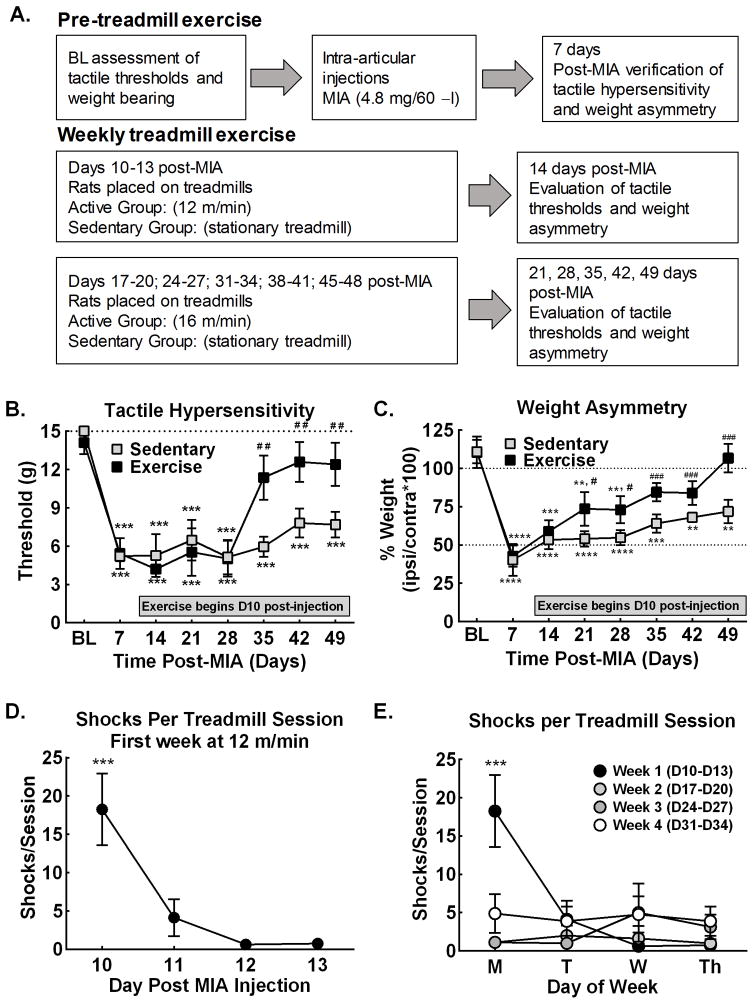
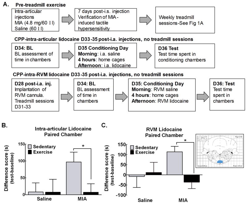
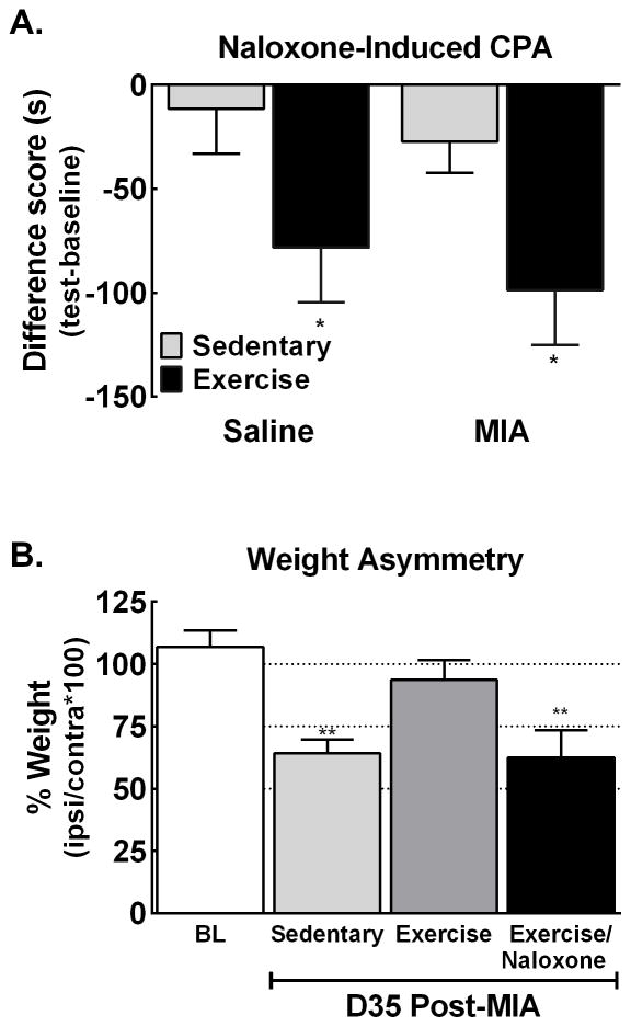
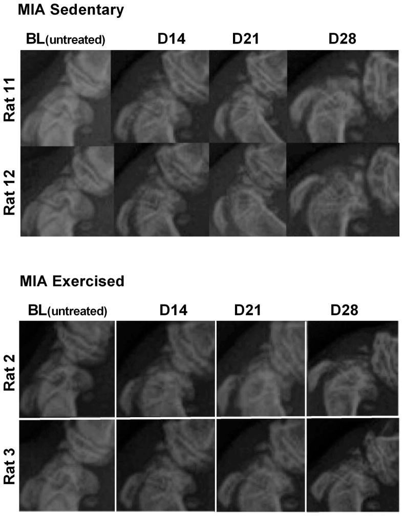
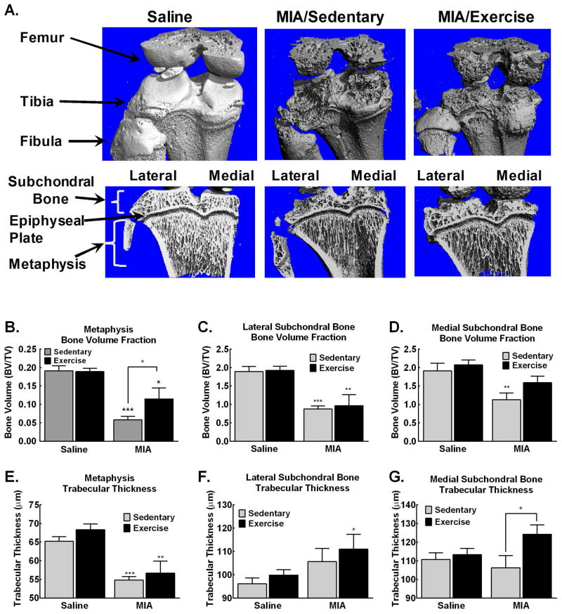
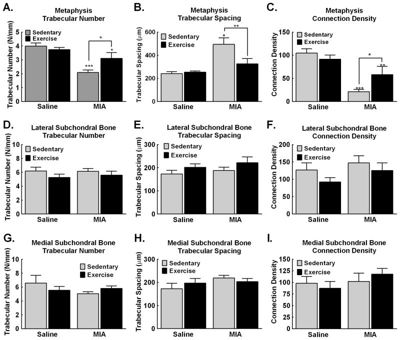
Methods: Rats treated with intraarticular (IA) monosodium iodoacetate (MIA) or saline underwent treadmill exercise or remained sedentary starting 10 days postinjection. Tactile sensory thresholds and weight bearing were assessed, followed by radiography at weekly intervals. After 4 weeks of exercise, ongoing pain was assessed using conditioned place preference (CPP) to IA or rostral ventromedial medulla (RVM)-administered lidocaine. The possible role of endogenous opioids in exercise-induced pain relief was examined by systemic administration of naloxone. Knee joints were collected for micro-computed tomography (micro-CT) analysis to examine pathologic changes to subchondral bone and metaphysis of the tibia.

Results: Treadmill exercise for 4 weeks reversed MIA-induced tactile hypersensitivity and weight asymmetry. Both IA and RVM lidocaine D35, administered post-MIA, induced CPP in sedentary but not exercised MIA-treated rats, indicating that exercise blocks MIA-induced ongoing pain. Naloxone reestablished weight asymmetry in MIA-treated rats undergoing exercise and induced conditioned place aversion, indicating that exercise-induced pain relief is dependent on endogenous opioids. Exercise did not alter radiographic evidence of OA. However, micro-CT analysis indicated that exercise did not block lateral subchondral bone loss or trabecular bone loss in the metaphysis, but did block MIA-induced medial bone loss.

Conclusion: These findings support the conclusion that exercise induces pain relief in advanced, NSAID-resistant OA, likely through increased endogenous opioid signaling. In addition, treadmill exercise blocked MIA-induced bone loss in this model, indicating a potential bone-stabilizing effect of exercise on the OA joint.


Conflict of interest statement

Conflict of interest: The authors have no conflict of interest

Figures

Figure 1
Figure 1. Treadmill exercise alleviates MIA-induced weight asymmetry and tactile hypersensitivity
A. Diagram of experimental procedure to examine effects of treadmill exercise on MIA-induced tactile hypersensitivity and weight asymmetry. B. Treadmill exercise starting 10 days post-MIA injection reversed tactile hypersensitivity selectively in exercise treated rats by D35 post-MIA, corresponding to 4 weeks of exercise. *p<0.05, **p<0.01, ***p<0.001 vs BL; ##p<0.01 vs Week 1. C. Treadmill exercise starting 10 days post-MIA injection into the knee joint reversed weight asymmetry in exercise, but not sedentary rats with improvement observed by D21 post-MIA, corresponding to 2 weeks of exercise, and continued improvement D35 through D49 post-MIA. *p<0.05, **p<0.01, ***p<0.001 vs BL, #p<0.05, ##p<0.01, ###p<0.001 vs Wk 1. D. Average number of shocks per treadmill session of MIA treated rats across the first week of treadmill exercise demonstrates that rats quickly adapt to running on the treadmill. Analysis or running across the 4-week treatment, ***p<0.05 vs D11, D12, and D13. E. Average number of shocks per daily treadmill session of MIA treated rats across the 4-week treatment demonstrates that the number of shocks were only elevated the first day of the first week of treadmill training. All graphs represent mean ± SEM.
Figure 2
Figure 2. Treadmill exercise alleviates MIA-induced ongoing pain
A. Diagram of experimental procedure to examine effects of treadmill exercise on MIA-induced ongoing pain using CPP to intra-articular or RVM lidocaine. B. Sedentary MIA treated rats significantly increased time spent in the intra-articular lidocaine paired chamber. Exercised rats treated with MIA failed to show an increase in time spent in the intra-articular lidocaine paired chamber (*p<0.05 vs sedentary MIA treated rats). Rats treated with intra-articular saline did not alter time spent in the intra-articular lidocaine paired chamber. C. Sedentary MIA treated significantly increased time spent in the chamber paired with RVM lidocaine. Exercised MIA treated rats failed to increase time spent in the RVM lidocaine paired chamber (*p<0.05 vs sedentary MIA treated rats). Intra-articular saline treated rats failed to alter time spent in the RVM lidocaine paired chamber irrespective of whether they were sedentary or exercised. All graphs represent mean ± SEM. Inset is a diagram illustrating verification of bilateral cannulation of the RVM. Map from The Rat Brain in Stereotaxic Coordinates, Paxinos and Watson, 1998. Highlighted area (gray) indicates region considered a hit. Ink from one or both needles outside this area were considered misses.
Figure 3
Figure 3. Treadmill exercise across 4 weeks starting 10-days post-injection increases opioid tone
A. MIA-induced weight asymmetry that persisted across 35 days in sedentary rats. Exercise starting 10 days post-MIA injection blocked the MIA-induced weight asymmetry. Naloxone (3 mg/kg i.p.) re-established the weight asymmetry at 30 min post-naloxone in the exercised rats. **p<0.01 vs BL, n=6 MIA sedentary, 5 MIA exercise. B. Analysis of difference scores confirms that exercised rats demonstrate equivalent decreases in time spent in the naloxone-paired chamber whereas sedentary rats failed to show significant CPA irrespective of whether they received intra-articular saline or intra-articular MIA. *p<0.05 vs sedentary rats. All graphs represent mean ± SEM.
Figure 4
Figure 4. Representative radiographs of sedentary or exercised MIA treated rats
Joint pathology was evident within 14 days post-MIA injection in both treatment groups. Radiographs indicate equivalent levels of MIA-induced joint pathology irrespective of whether the rats remained sedentary or underwent exercise.
Figure 5
Figure 5. MIA-induced pathological changes in the subchondral and metaphysis are blocked by treadmill exercise
A. Representative images demonstrate MIA-induced bone remodeling of the exterior bone, with development of osteophytes and bone deformities. Trabecular bone loss is observed within subchondral bone and metaphysis. B. Significant reduction in bone volume is observed in the medial subchondral bone of MIA treated sedentary, but not MIA treated exercise rats. C. Significant reduction in bone volume is observed in the lateral subchondral bone of MIA treated sedentary and exercise treated rats. D. Diminished bone volume is observed in the trabecular region of the metaphysis in the MIA treated sedentary rats that is attenuated in the MIA treated rats that underwent treadmill exercise. E. Significant reduction of trabecular bone thickness is observed in the metaphysis of in MIA treated rats. F. Increased trabecular thickness is observed in the lateral subchondral bone of MIA treated rats. G. Increased trabecular thickness within the medial subchondral bone is observed in MIA treated rats that underwent exercise across 4 weeks. *p<0.05, **p<0.01, ***p<0.001 vs saline sedentary.
Figure 6
Figure 6. MicroCT analysis of the tibia demonstrates that MIA-induced pathological changes in the subchondral and metaphysis are blocked by treadmill exercise
A. Analysis of the number of trabeculae within the metaphysis demonstrates decreased trabeculae in MIA treated sedentary rats. Exercise blocked the MIA-induced decrease in trabecular number, but the number was still significantly lower compared to saline sedentary control rats. B. Analysis of trabecular spacing demonstrates increased space between trabecular bone within the metaphysis in the sedentary MIA treated rats. Exercise blocked the MIA-induced increase in trabecular spacing, with trabecular spacing similar to saline sedentary control rats. C. MIA reduced connection density of trabecular bone within the metaphysis. Exercise attenuated the MIA induced reduction in connection density of trabecular bone. D–F. MIA and exercise both failed to alter the number (D), spacing (E) or connection density (F) of the trabecular bone within the lateral subchondral bone. G–I. MIA and exercise both failed to alter the number (G), spacing (H) or connection density (I) of the trabecular bone within the medial subchondral bone. Graphs represent mean ± SEM. *p<0.05, **p<0.01, ***p<0.001 vs saline sedentary.

References

    1. Dillon CF, Rasch EK, Gu Q, Hirsch R. Prevalence of knee osteoarthritis in the United States: arthritis data from the Third National Health and Nutrition Examination Survey 1991–94. J Rheumatol. 2006;33(11):2271–9. - PubMed
    1. Hugle T, Geurts J, Nuesch C, Muller-Gerbl M, Valderrabano V. Aging and Osteoarthritis: An Inevitable Encounter? Journal of Aging Research. 2012;2012:7. - PMC - PubMed
    1. Hunter DJ, McDougall JJ, Keefe FJ. The symptoms of OA and the genesis of pain. Rheumatic diseases clinics of North America. 2008;34(3):623–43. - PMC - PubMed
    1. Mobasheri A, Batt M. An update on the pathophysiology of osteoarthritis. Annals of Physical and Rehabilitation Medicine. 2016;59(5–6):333–9. - PubMed
    1. Neogi T, Zhang Y. Epidemiology of OA. Rheumatic diseases clinics of North America. 2013;39(1):1–19. - PMC - PubMed

Publication types

MeSH terms