. 2018 Jan;175(2):272-283.
doi: 10.1111/bph.13790. Epub 2017 May 12.

Dexpramipexole improves bioenergetics and outcome in experimental stroke

Affiliations

Dexpramipexole improves bioenergetics and outcome in experimental stroke

Mirko Muzzi et al. Br J Pharmacol. 2018 Jan.

Abstract

Background and purpose: Dexpramipexole, a drug recently tested in patients with amyotrophic lateral sclerosis (ALS,) is able to bind F1Fo ATP synthase and increase mitochondrial ATP production. Here, we have investigated its effects on experimental ischaemic brain injury.

Experimental approach: The effects of dexpramipexole on bioenergetics, Ca2+ fluxes, electrophysiological functions and death were evaluated in primary neural cultures and hippocampal slices exposed to oxygen-glucose deprivation (OGD). Effects on infarct volumes and neurological functions were also evaluated in mice following proximal or distal middle cerebral artery occlusion (MCAo). Distribution of dexpramipexole within the ischaemic brain was evaluated by means of mass spectrometry imaging.

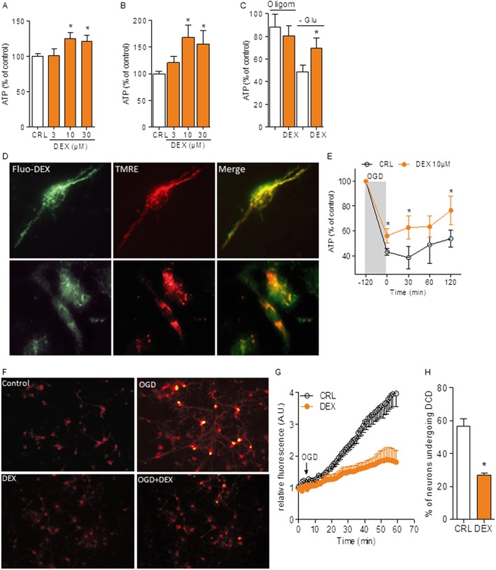
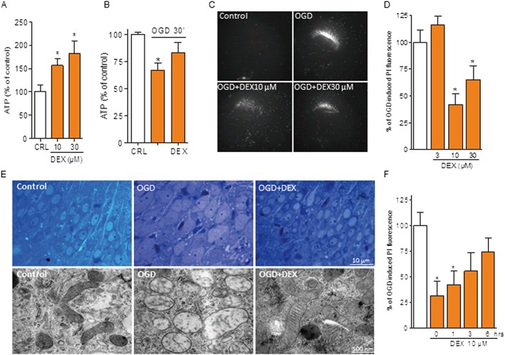
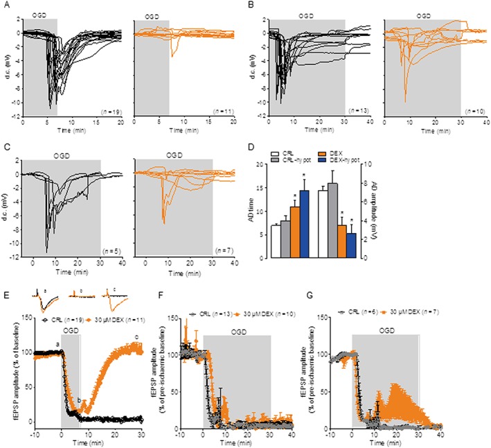
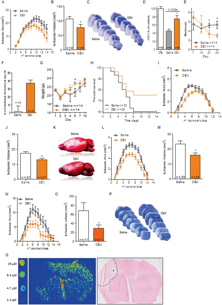
Key results: Dexpramipexole increased mitochondrial ATP production in cultured neurons or glia and reduces energy failure, prevents intracellular Ca2+ overload and affords cytoprotection when cultures are exposed to OGD. This compound also counteracted ATP depletion, mitochondrial swelling, anoxic depolarization, loss of synaptic activity and neuronal death in hippocampal slices subjected to OGD. Post-ischaemic treatment with dexpramipexole, at doses consistent with those already used in ALS patients, reduced brain infarct size and ameliorated neuroscore in mice subjected to transient or permanent MCAo. Notably, the concentrations of dexpramipexole reached within the ischaemic penumbra equalled those found neuroprotective in vitro.

Conclusion and implications: Dexpramipexole, a compound able to increase mitochondrial F1Fo ATP-synthase activity reduced ischaemic brain injury. These findings, together with the excellent brain penetration and favourable safety profile in humans, make dexpramipexole a drug with realistic translational potential for the treatment of stroke.

Linked articles: This article is part of a themed section on Inventing New Therapies Without Reinventing the Wheel: The Power of Drug Repurposing. To view the other articles in this section visit http://onlinelibrary.wiley.com/doi/10.1111/bph.v175.2/issuetoc.


Figures

Figure 1
Figure 1
DEX improves neural cell bioenergetics and resistance to OGD. ATP contents in resting mouse cultures of pure cortical neurons (A) or glial cells (B) after a 6 h exposure to different DEX concentrations. Values of ATP under control condition were 8.3 ± 2.21 and 1.1 ± 0.54 nmol·mg protein−1 for neurons and glia respectively. (C) Effects of the F1Fo ATP synthase inhibitor oligomycin (10 µM) or glucose starvation in the presence of pyruvate and glutamine on ATP content in glial cells exposed to DEX (10 µM). (D) Representative images of the intracellular distribution of 10 µM Fluo‐DEX (green) in the absence or presence of unlabelled 100 µM DEX and the potentiometric mitochondrial dye TMRE (red) in hippocampal neurons in culture. (E) ATP contents in cultures of mixed cortical cells during OGD and after returning to normoxia and normal growth medium. Value of ATP under control condition was 10.5 ± 1.82 nmol·mg protein−1. (F) Visualization of intracellular Ca2+ contents in primary cultures of hippocampal neurons exposed to OGD. DEX was added to the culture 10 min before OGD. The temporal pattern of Ca2+ increase (G) and percentage of neurons undergoing DCD (H) are also shown. Data represent the mean ± SEM of at least three experiments. *P < 0.05, significantly different from corresponding control (CRL); ANOVA plus Tukey's post hoc test.
Figure 2
Figure 2
DEX protects hippocampal slices from OGD‐dependent neurodegeneration. ATP content of organotypic hippocampal slices 6 h after different concentrations of DEX (A) or immediately after OGD (30 min) in the absence or presence of 10 µM DEX added 10 min before OGD (B). Value of ATP under control condition was 14.8 ± 3.51 nmol·mg protein−1. Representative images (C) and quantification (D) of the effect of different concentrations of DEX (added at the end of the OGD insult) on cell death (revealed by PI staining) of CA1 neurons of organotypic hippocampal slices exposed to 30 min OGD/24 h reoxygenation. (E) DEX (10 µM 10 min before OGD) prevents swelling of somata of hippocampal CA1 neurons and their mitochondria in organotypic hippocampal slices exposed to 30 min OGD. (F) Effect of different post‐treatment windows with 10 µM DEX on cell death (revealed by PI staining) of CA1 neurons of organotypic hippocampal slices exposed to 30 min OGD/24 h reoxygenation. Data represent the mean ± SEM of at least three experiments. *P < 0.05, significantly different from corresponding control (CRL); ANOVA plus Tukey's post hoc test.
Figure 3
Figure 3
DEX protects hippocampal slices from OGD‐dependent anoxic depolarization. (A) AD (expressed as d.c. shift) occurring in acute hippocampal slices exposed to 7 min OGD is prevented by co‐incubation with DEX (30 µM, red traces). (B) AD occurring in acute hippocampal slices exposed to 30 min OGD is not prevented by co‐incubation with DEX (30 µM). (C) DEX suffices to prevent AD in three out of seven slices exposed to 30 min OGD under hypothermic conditions (30°C bath temperature). (D) Effect of 30 µM DEX on AD time and amplitude in slices exposed to 30 min OGD under normothermic or hypothermic (30°C) conditions. (E) Neurotransmission (measured as filed EPSP) in the CA1 region of acute hippocampal slices exposed to 7 min OGD is irreversibly lost in control slices but fully restored upon reoxygenation in those incubated with DEX 30 µM. DEX does not prevent loss of neurotransmission in slices exposed to 30 min OGD (F) but leads to partial and transient recovery under hypothermic conditions (30°C) (G). Data represent the mean ± SEM of at least three experiments. *P < 0.05, significantly different from corresponding control (CRL); ANOVA plus Tukey's post hoc test.
Figure 4
Figure 4
DEX is functionally neuroprotective in experimental stroke models. DEX (3 mg·kg−1 i.p.) administered at reperfusion and then every 12 h reduces infarct areas (A) and volumes (B) of mice subjected to 1 h MCAo/48 h reperfusion. (C) Visualization of infarct distribution (pale areas) in toluidine blue‐stained coronal brain slices of representative saline‐ or DEX‐treated mice. (D) Content of ATP in the brain of control mice (10.3 ± 4.06 nmol·mg protein−1) and in the penumbra of ischaemic (1 h MCAo) brain 3 h after reperfusion in mice treated or not with DEX 3 mg·kg−1 i.p. Effect of a 7 day treatment with DEX (3 mg·kg−1 i.p. bid) on neuroscore (E) and functional recovery (F) over 1 month in mice subjected to 1 h MCAo. Effect of DEX on weight recovery (G) and survival (H) compared with saline treatment in mice subjected to 1 h MCAo. Effect of DEX (3 mg·kg−1 i.p. bid, first dose immediately after artery occlusion) on infarct areas (I) and volumes (J) of mice subjected to permanent MCAo (distal MCA cauterization). (K) Visualization by TTC staining of the extent of cortical infarct (pale area) in representative brains of saline‐ or DEX‐treated mice. Effect of a 1 h post‐treatment with DEX (3 mg·kg−1 bid) on cortical infarct areas (L) or volumes (M) of mice subjected to permanent MCAo. Effect of DEX (3 mg·kg−1 i.p. bid, first dose immediately after artery occlusion) on infarct areas (N) and volumes (O) of rats subjected to permanent MCAo. (P) Visualization of infarct distribution (pale areas) in toluidine blue‐stained coronal brain slices of representative saline‐ or DEX‐treated rats. (Q) Pseudocolour visualization by means of imaging mass spectrometry of DEX distribution in a mouse brain subjected to 48 h distal MCAo cauterization and 1 h treatment with 3 mg·kg−1 DEX i.p. The pseudocolour intensity of standard concentrations spotted on a control brain slice is shown on the left. Note that the increased signal in the slice fracture is an artefact due to analyte delocalization (see Methods). Haematoxylin and eosin staining of the slice analysed by mass spectrometry is shown on the right. The dotted line delimits the infarct boundary. Data represent the mean ± SEM of at least three experiments. *P < 0.05, significantly different from saline; Student's t‐test or ANOVA plus Tukey's post hoc test.

Similar articles

Cited by

References

    1. Alavian KN, Dworetzky SI, Bonanni L, Zhang P, Sacchetti S, Mariggio MA et al. (2012). Effects of dexpramipexole on brain mitochondrial conductances and cellular bioenergetic efficiency. Brain Res 1446: 1–11. - PMC - PubMed
    1. Alavian KN, Dworetzky SI, Bonanni L, Zhang P, Sacchetti S, Li H et al. (2015). The mitochondrial complex V‐associated large‐conductance inner membrane current is regulated by cyclosporine and dexpramipexole. Mol Pharmacol 87: 1–8. - PMC - PubMed
    1. Alexander SP, Kelly E, Marrion N, Peters JA, Benson HE, Faccenda E et al. (2015a). The Concise Guide to PHARMACOLOGY 2015/16: Transporters. Br J Pharmacol 172: 6110–6202. - PMC - PubMed
    1. Alexander SPH, Davenport AP, Kelly E, Marrion N, Peters JA, Benson HE et al. (2015b). The Concise Guide to PHARMACOLOGY 2015/16: G protein‐coupled receptors. Br J Pharmacol 172: 5744–5869. - PMC - PubMed
    1. Bernardi P, Rasola A, Forte M, Lippe G (2015). The mitochondrial permeability transition pore: channel formation by F‐ATP synthase, integration in signal transduction, and role in pathophysiology. Physiol Rev 95: 1111–1155. - PMC - PubMed

Publication types

MeSH terms