Review
. 2017 Apr 15:375:8-17.
doi: 10.1016/j.jns.2017.01.033. Epub 2017 Jan 10.

Acquired pendular nystagmus

Affiliations
Review

Acquired pendular nystagmus

Sarah Kang et al. J Neurol Sci. .

Abstract

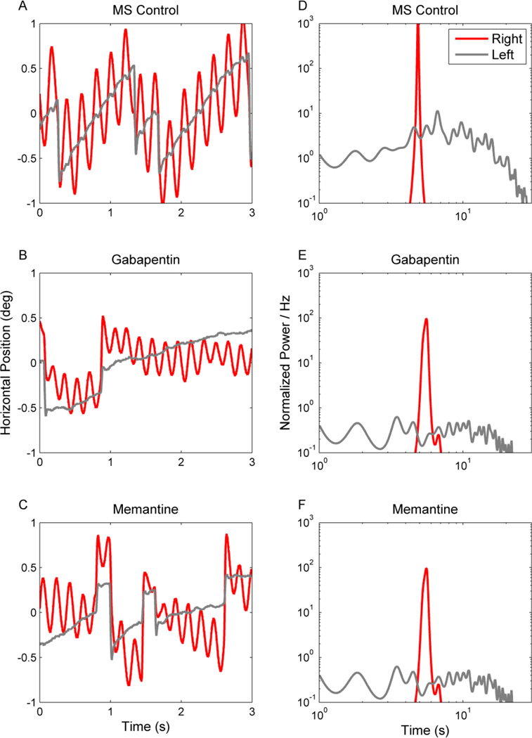
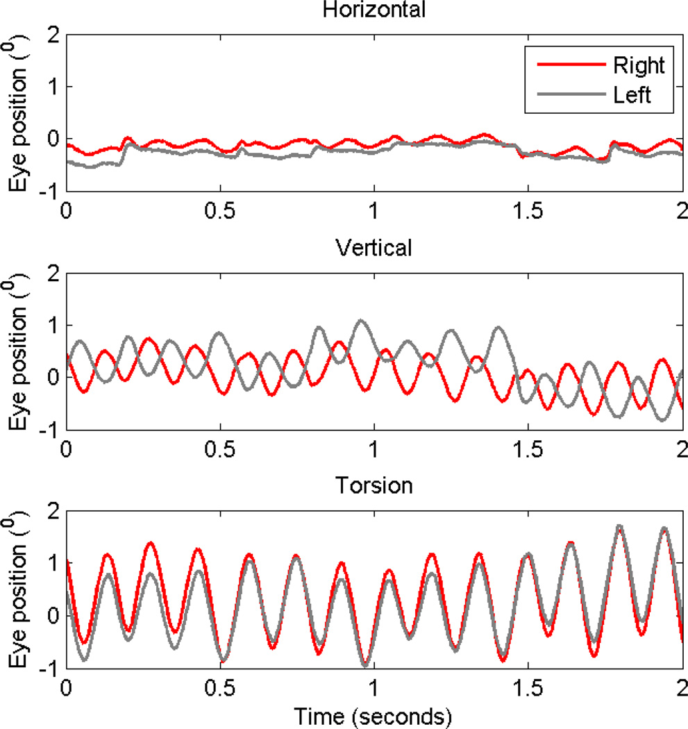
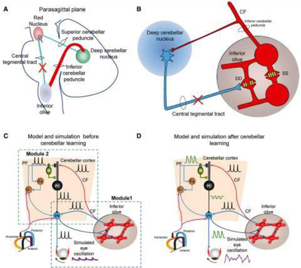
Acquired pendular nystagmus is comprised of quasi-sinusoidal oscillations of the eyes significantly affecting gaze holding and clarity of vision. The most common causes of acquired pendular nystagmus include demyelinating disorders such as multiple sclerosis and the syndrome of ocular palatal tremor. However, several other deficits, such as pharmacological intoxication, metabolic and genetic disorders, and granulomatous disorders can lead to syndromes mimicking acquired pendular nystagmus. Study of the kinematic features of acquired pendular nystagmus has suggested a putative pathophysiology of an otherwise mysterious neurological disorder. Here we review clinical features of neurological deficits that co-occur with acquired pendular nystagmus. Subsequent discussion of the pathophysiology of individual forms of pendular nystagmus speculates on mechanisms of the underlying disease while providing insights into pharmacotherapy of nystagmus.

Keywords: Degenerative disorder; Demyelination disorder; Multiple sclerosis; Stroke; Toxin exposure.


Figures

Figure 1
Figure 1
Schematic function of and organization of the neural integrator and critical feedback loops.
Figure 2
Figure 2
Horizontal acquired pendular nystagmus measured from one patient with MS. (A–C) Binocular eye positions are plotted against time. Red traces depict the right eye and grey traces the left. IN this example binocular jerk nystagmus superimposes upon monocular acquired pendular nystagmus in the right eye. There is a reduction in the amplitude of both types of nystagmus during treatment with gabapentin (1200 mg per day) (B) and memantine (10 mg per day) (C). Corresponding power spectra are plotted in D–F. The power spectrum of the pendular component is very sharp (D, red trace). The power spectrum of the jerk nystagmus is broader and lower (D, gray trace). Note that both gabapentin and memantine reduce the oscillation amplitude, but do not significantly affect its frequency distribution.
Figure 3
Figure 3
This is an example of acquired pendular nystagmus in a subject with sarcoidosis. Horizontal (A), vertical (B) and torsional (C) eye positions are plotted against time. Red traces are right eye positions, while grey traces are left. Horizontal oscillations have minimal amplitude, while the amplitude increased in vertical and then in torsional direction. Torsional oscillations are conjugate, while vertical oscillations are disconjugate as depicted by 180° phase shift.
Figure 4
Figure 4
Schematic representation of the Guillain–Mollaret triangle that is comprised of fibers connecting the deep cerebellar nuclei and contralateral inferior olive. These fibers pass near the red nucleus without any rubral relay (A). The coupling strength through the connexn gap junctions (schematized with yellow channels; DD) between neghboring inferior olivary neurons are inhibited by projections from the deep cerebellar nuclei (blue projection) (B). Lesions in the Guillain–Mollaret triangle (red X in A and B) leads to hypertrophy of the inferior olive and subsequently increased soma-somatic gap junction. Schematic representation of a “dual-mechanism model” (C and D). Model and traces from simulations after inferior olive hypertrophy but before maladaptive cerebellar modulation (C). Inferior olive and cerebellar modules after hypertrophy and maladaptation of the cerebellum module (D). Lower left corner shows icon for semicircular canals (C and D). Simulated membrane potentials (black), eye oscillations (magenta). CF = climbing fibres; PF = parallel fibres; DD = dendro-dendritic gap junction; SS = soma-somatic gap junction; Gr = granule cell layer; IN = interneurons; PC = Purkinje neurons.
Figure 5
Figure 5
This is an example of the attenuation of horizontal acquired pendular nystagmus by gabapentin and memantine in an OPT patient. (A–C) Binocular eye positions are plotted against time. Red traces represent the right eye positions while grey lines are left. The oscillations are irregular and disconjugate. The amplitude is reduced during treatment with both gabapentin (B) and memantine (C). Corresponding power spectra are plotted in D–F. The power spectrum is very broad, with a large peak around 2 Hz. Gabapentin and memantine not only reduce the oscillation amplitude but they also reduce the irregularity of the waveform as depicted by the reduction of the range of its frequency distribution.

Similar articles

Cited by

References

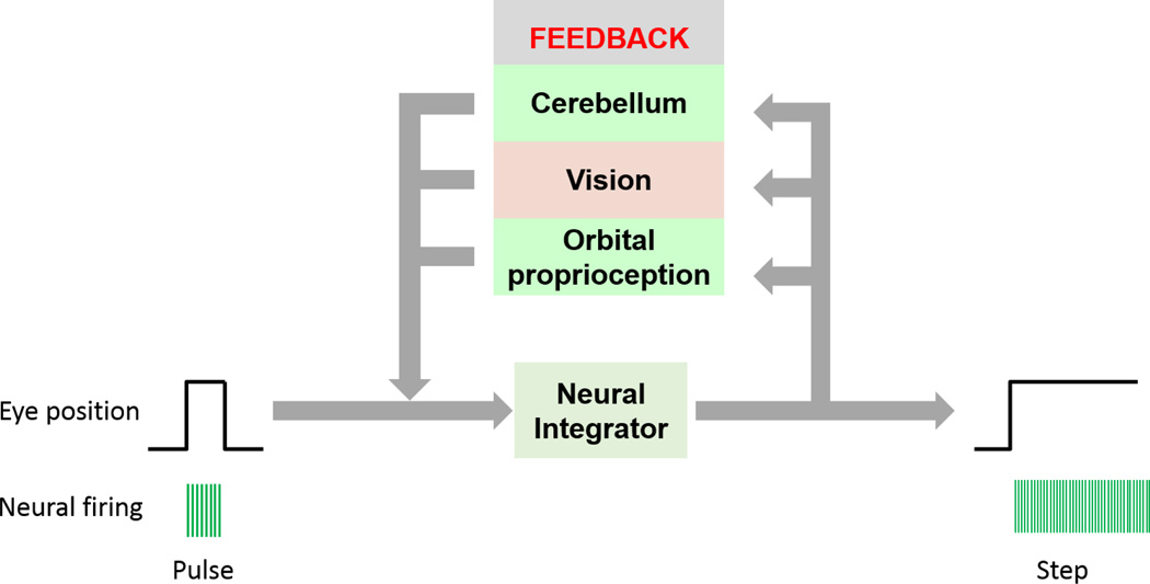
    1. Cannon SC, Robinson DA. An improved neural-network model for the neural integrator of the oculomotor system: more realistic neuron behavior. Biological cybernetics. 1985;53:93–108. - PubMed
    1. Cannon SC, Robinson DA. Loss of the neural integrator of the oculomotor system from brain stem lesions in monkey. Journal of neurophysiology. 1987;57:1383–1409. - PubMed
    1. Cannon SC, Robinson DA, Shamma S. A proposed neural network for the integrator of the oculomotor system. Biological cybernetics. 1983;49:127–136. - PubMed
    1. Robinson DA. The effect of cerebellectomy on the cat’s bestibulo-ocular integrator. Brain research. 1974;71:195–207. - PubMed
    1. Frohman EM, Frohman TC, Zee DS, McColl R, Galetta S. The neuro-ophthalmology of multiple sclerosis. The Lancet Neurology. 2005;4:111–121. - PubMed

MeSH terms