. 2017 Apr 3;216(4):943-960.
doi: 10.1083/jcb.201610108. Epub 2017 Mar 20.

Molecular mechanism of dynein recruitment to kinetochores by the Rod-Zw10-Zwilch complex and Spindly

Affiliations

Molecular mechanism of dynein recruitment to kinetochores by the Rod-Zw10-Zwilch complex and Spindly

José B Gama et al. J Cell Biol. .

Abstract

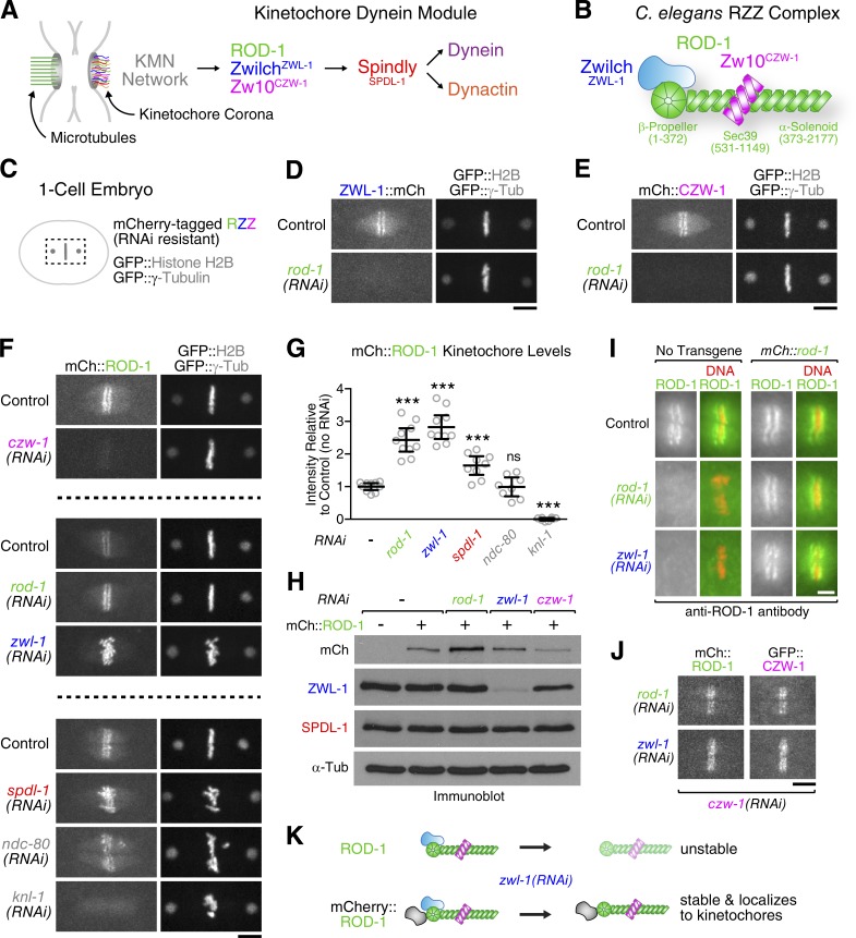
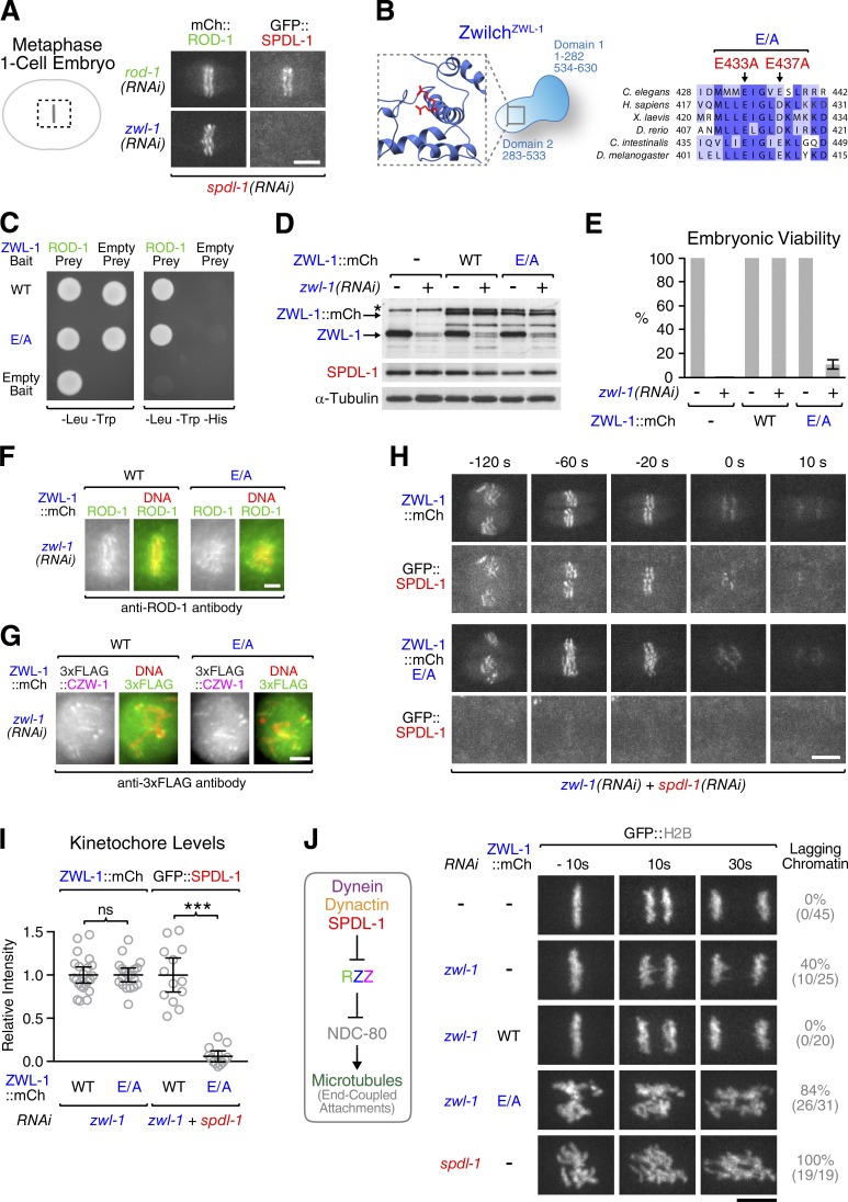
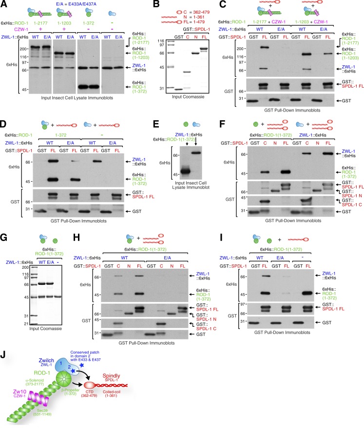
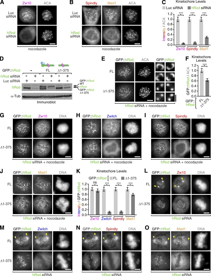
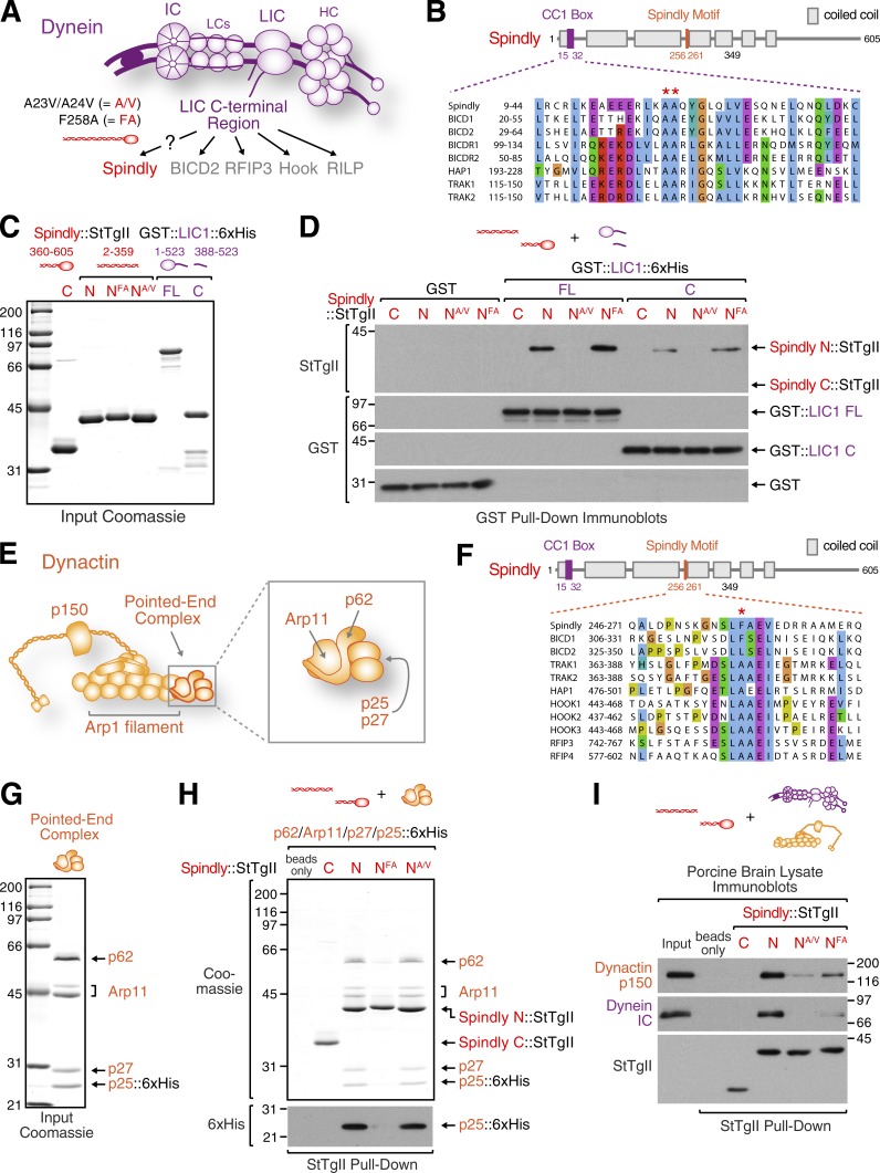
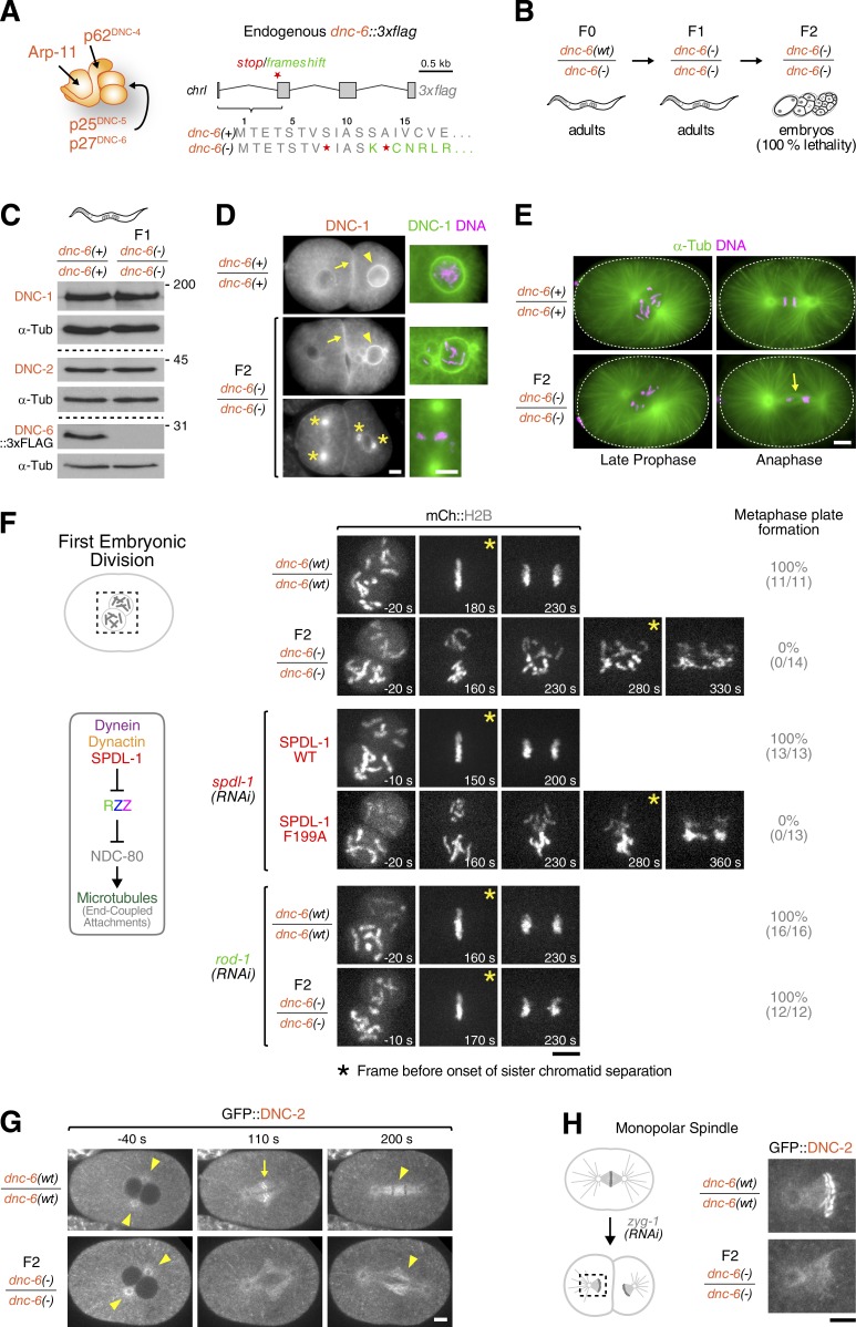
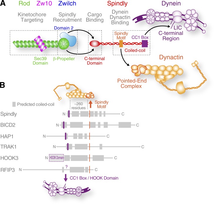
The molecular motor dynein concentrates at the kinetochore region of mitotic chromosomes in animals to accelerate spindle microtubule capture and to control spindle checkpoint signaling. In this study, we describe the molecular mechanism used by the Rod-Zw10-Zwilch complex and the adaptor Spindly to recruit dynein to kinetochores in Caenorhabditis elegans embryos and human cells. We show that Rod's N-terminal β-propeller and the associated Zwilch subunit bind Spindly's C-terminal domain, and we identify a specific Zwilch mutant that abrogates Spindly and dynein recruitment in vivo and Spindly binding to a Rod β-propeller-Zwilch complex in vitro. Spindly's N-terminal coiled-coil uses distinct motifs to bind dynein light intermediate chain and the pointed-end complex of dynactin. Mutations in these motifs inhibit assembly of a dynein-dynactin-Spindly complex, and a null mutant of the dynactin pointed-end subunit p27 prevents kinetochore recruitment of dynein-dynactin without affecting other mitotic functions of the motor. Conservation of Spindly-like motifs in adaptors involved in intracellular transport suggests a common mechanism for linking dynein to cargo.

Keywords: mitosis; kinetochore; dynein/dynactin; BICD2; Spindly; RZZ complex.


Figures

Figure 1.
Figure 1.
ROD-1 and Zw10CZW-1 can target to kinetochores independently of ZwilchZWL-1. (A) Cartoon depicting localization dependencies of kinetochore dynein module components. (B) Summary of interactions between C. elegans RZZ subunits, based on yeast two-hybrid mapping (Fig. S1, A–C). Note that the intact RZZ complex likely exists as a dimer of the ROD-1–Zw10CZW-1–ZwilchZWL-1 trimer, based on hydrodynamic analysis of the human and D. melanogaster complexes (Williams et al., 2003; Çivril et al., 2010). (C) Cartoon of the one-cell embryo spindle region shown in subsequent panels. (D–F) Localization dependency analysis of mCherry-tagged RZZ subunits. Still images are from time-lapse sequences. (G) Quantification of mCherry::ROD-1 levels at kinetochores in the conditions shown in D–F. Circles correspond to measurements in individual one-cell embryos in metaphase. Error bars represent the SEM with a 95% confidence interval. The t test was used to determine statistical significance (***, P < 0.0001 compared with no RNAi control; ns, P > 0.05). (H) Immunoblots showing that depletion of Zw10CZW-1 but not ZwilchZWL-1 lowers mCherry::ROD-1 levels. α-Tubulin served as the loading control. (I) Immunofluorescence images of metaphase kinetochores stained for ROD-1. Endogenous ROD-1 is missing from kinetochores in ZwilchZWL-1-depleted embryos, whereas mCherry::ROD-1 localizes under the same conditions. (J) Stills from a time-lapse sequence showing that mCherry::ROD-1 supports kinetochore localization of GFP::Zw10CZW-1 in the absence of ZwilchZWL-1. Bars: (D–F and J) 5 µm; (I) 2µm. (K) Cartoon summary of the data shown in C–J.
Figure 2.
Figure 2.
ZwilchZWL-1 residues required for SpindlySPDL-1 recruitment to kinetochores. (A) Stills from time-lapse sequences showing that kinetochore-localized mCherry::ROD-1 does not support GFP::SpindlySPDL-1 recruitment in the absence of ZwilchZWL-1. (B) Cartoon of domain architecture and sequence alignment of Zwilch homologues. Residues E433 and E437 were both mutated to alanine (E/A). (C) Yeast two-hybrid assay showing that the ZwilchZWL-1 E/A mutant interacts with full-length ROD-1. Cells containing bait and prey plasmids grow on -Leu/-Trp plates, whereas -Leu/-Trp/-His plates select for bait–prey interaction. (D) Immunoblots of C. elegans adult worms expressing transgene-encoded ZwilchZWL-1::mCherry wild-type (WT) or the E/A mutant. The asterisk denotes a cross-reacting protein band of similar size as ZwilchZWL-1::mCherry. α-Tubulin served as the loading control. (E) Embryonic viability assay. More than 300 embryos from 12 or more mothers were counted for each condition. (F and G) Still images from a time-lapse sequence showing robust kinetochore localization of ROD-1 and 3×FLAG::Zw10CZW-1 in the ZwilchZWL-1 E/A mutant. (H) Selected frames from a time-lapse sequence showing that ZwilchZWL-1::mCherry E/A fails to recruit GFP::SpindlySPDL-1 (see Video 1). (I) Quantification of ZwilchZWL-1::mCherry and GFP::SpindlySPDL-1 levels on metaphase kinetochores in the conditions shown in H. Circles correspond to measurements in individual embryos. Error bars represent the SEM with a 95% confidence interval. The t test was used to determine statistical significance (***, P < 0.0001; ns, P > 0.05). (J, left) Cartoon summarizing the inhibitory cross-talk between the kinetochore dynein module and NDC-80. (Right) Selected frames from time-lapse sequences of the first embryonic division, demonstrating that the chromosome segregation defects of ZwilchZWL-1::mCherry E/A resemble those of SpindlySPDL-1 depletion (see Video 2). The frequencies of anaphase chromatin bridges are indicated in the percentages and absolute numbers of embryos in parentheses. Time is relative to the onset of sister chromatid separation. Bars: (A, H, and J) 5 µm; (F and G) 2 µm.
Figure 3.
Figure 3.
SpindlySPDL-1 binds ZwilchZWL-1 and the ROD-1 β-propeller. (A) Lysates of insect Sf21 cells coinfected with viruses to express full-length (FL) ROD-11–2,177ZZ, ROD-11–1,203ZZ, ROD-11–372–ZwilchZWL-1, or ZwilchZWL-1 alone. ROD-1 and ZwilchZWL-1 were tagged with 6×His for detection on immunoblots. (B) Coomassie-stained protein gels showing purified recombinant GST::SpindlySPDL-1 used in pull-downs. (C and D) GST pull-downs from the lysates in A with purified GST::SpindlySPDL-1 from B. Protein fractions bound to beads were analyzed by immunoblotting using anti-6×His antibody. The same membranes were then reprobed with anti-GST antibody. (E) Lysates of insect Sf21 cells infected with viruses encoding for the ROD-1 β-propeller (residues 1–372) or ZwilchZWL-1, both tagged with 6×His. (F) Immunoblots of GST pull-downs from the lysates in E with purified GST::SpindlySPDL-1 (see A). (G) Coomassie-stained protein gels showing purified ROD-1 β-propeller alone and in complex with wild-type (WT) ZwilchZWL-1 or the E/A mutant. (H and I) Immunoblots of GST pull-downs using the purified proteins in B and G. (J) Cartoon showing the bipartite interaction between RZZ and the C-terminal domain (CTD) of SpindlySPDL-1. Molecular mass is indicated in kilodaltons.
Figure 4.
Figure 4.
Human Rod lacking its β-propeller localizes to kinetochores with Zw10 but fails to recruit Zwilch or Spindly. (A and B) HeLa cells coimmunostained with ACAs and Spindly/Mad1 (A) or Zw10 (B) after transfection with an siRNA oligonucleotide against hRod or luciferase (Luc) as a control. Cells were incubated in 1 µM nocodazole for 4 h before fixation. (C) Quantification of kinetochore levels of Spindly, Mad1, and Zw10 using immunofluorescence intensity measurements normalized to ACA signal. Each condition represents ≥50 kinetochore measurements from 10 or more different cells. Error bars represent the SEM with a 95% confidence interval. The t test was used to determine statistical significance (***, P < 0.0001). (D) Immunoblot of HeLa Flp-In T-REx cells with an antibody against human Rod (hRod), showing expression levels of endogenous hRod and RNAi-resistant full-length GFP::hRod (FL) or GFP::hRod lacking the β-propeller domain (Δ1–375). Luciferase siRNA was used as a control in RNAi experiments, and α-tubulin served as the loading control. Note that GFP::hRod(Δ1–375) migrates at the same size as endogenous hRod. (E) HeLa Flp-In T-REx cells expressing GFP::hRod or GFP::hRod(Δ1–375), depleted of endogenous hRod and immunostained with anti-GFP and ACAs. Blow-ups show examples of individual sister kinetochore pairs. (F) Quantification of full-length and Δ1–375 GFP::hRod levels at kinetochores as in C. (G–J) HeLa Flp-In T-REx cells immunostained for GFP and Zw10 (G), Zwilch (H), Spindly (I), or Mad1 (J) in cells expressing full-length and Δ1–375 GFP::hRod after depletion of endogenous hRod. (K) Quantification of kinetochore levels for the components in G–J as described in C using immunofluorescence intensity measurements normalized to GFP::hRod signal. (L–O) HeLa Flp-In T-REx cells in prometaphase immunostained as in G–J but without nocodazole treatment. Arrowheads point to the accumulation of GFP::hRod at spindle poles along with Zw10, Zwilch, Spindly, and Mad1. Polar accumulation is missing in cells expressing GFP::hRod(Δ1–375). Bars: (A, B, E [main images], G–J, and L–O) 5 µm; (E, zoom) 1 µm.
Figure 5.
Figure 5.
Spindly binds dynein LIC and the dynactin pointed-end complex. (A) Cartoon of dynein, composed of two heavy chains (HCs), two ICs, two LICs, and six light chains (LCs). The C-terminal region of LIC binds dynein adaptors involved in membrane transport. The two point mutants of human Spindly used throughout this figure are indicated. (B) Sequence alignment showing a conserved region in the first coiled-coil segment of Spindly and other dynein adaptors (CC1 box). The two alanines mutated to valine in Spindly (A23/A24) and BICD2 (A43/A44) are marked with asterisks. (C) Coomassie-stained gels of purified Spindly fragments fused to Strep-tag II (StTgII) as well as GST-tagged full-length (FL) and C-terminal LIC1. (D) Immunoblots of GST pull-downs using the proteins in C. Protein fractions bound to beads were detected on blots with Strep-Tactin. The same membrane was then reprobed with anti-GST antibody. (E) Cartoon of dynactin with the pointed-end complex highlighted. (F) Sequence alignment showing that several dynein adaptors possess a motif that in Spindly is implicated in dynein–dynactin recruitment to kinetochores. The phenylalanine mutated to alanine in Spindly (F258) is marked with an asterisk. (G) Coomassie-stained gels of the purified recombinant dynactin pointed-end complex. The p25 subunit is tagged with 6×His. Arp11 appears as two distinct bands. We verified that both bands correspond to Arp11 by expressing Arp11::6×His (unpublished data). (H) Coomassie-stained gel and immunoblot of Strep-tag II pull-downs using the purified proteins in C and G. (I) Immunoblots of Strep-tag II pull-downs from porcine brain lysate using the purified proteins in C. The same membrane was probed for dynactin p150, dynein IC, and Strep-tagged Spindly. Molecular mass is indicated in kilodaltons.
Figure 6.
Figure 6.
Dynactin p27DNC-6 is required for dynein–dynactin recruitment to kinetochores. (A, left) Cartoon of the C. elegans dynactin pointed-end complex. (Right) Schematic of the endogenous p27dnc-6 locus modified with a C-terminal 3×flag tag. Stop codons (red stars) and a frameshift mutation (green lettering) were subsequently introduced to create a null allele, dnc-6(−). (B) Genetics of the p27dnc-6-null allele: F0 mothers heterozygous for dnc-6(−) contain a balancer chromosome with a wild-type copy, dnc-6(wt). F1 progeny homozygous for dnc-6(−) that survive to adulthood on maternally provided p27DNC-6 produce F2 dnc-6(−/−) embryos that completely lack p27DNC-6. (C) Immunoblots of F1 adults homozygous for dnc-6(−), demonstrating that levels of dynactin p150DNC-1 and p50DNC-2 are unaffected by the absence of p27DNC-6. Molecular mass is indicated in kilodaltons. (D) Immunofluorescence images of F2 dnc-6(−/−) embryos at the two-cell stage stained with anti-p150DNC-1 antibody, showing that dynactin without p27DNC-6 is recruited to the nuclear envelope (arrowheads), the cell cortex (arrows), and centrosomes (asterisks). Blowups show evidence of chromosome missegregation and aneuploidy in F2 dnc-6(−/−) embryos. (E) Immunofluorescence images showing successful bipolar spindle assembly in F2 dnc-6(−/−) embryos. The arrow indicates chromosome missegregation. (F) Selected time-lapse sequence frames of the first mitotic division in embryos expressing mCherry::histone H2B. F2 dnc-6(−/−) embryos exhibit severe chromosome segregation defects that are similar to those observed for the SpindlySPDL-1 F199A mutant. The defects are significantly ameliorated after depletion of ROD-1, as predicted by the inhibitory cross-talk between the kinetochore dynein module and NDC-80 (summarized in schematic on the left). The frequencies of embryos in which chromosomes congress to metaphase plates are indicated in the percentages and absolute numbers of embryos in parentheses. Time is relative to nuclear envelope breakdown. (G) Selected frames from a time-lapse sequence in F2 dnc-6(−/−) embryos expressing GFP::p50DNC-2, demonstrating that dynactin without p27DNC-6 localizes normally to centrosomes and the mitotic spindle (arrowheads) but is absent from kinetochores (arrow). (H, left) Experimental strategy to generate monopolar spindles in F2 dnc-6(−/−) embryos expressing GFP::p50DNC-2 by depleting the centriole duplication kinase ZYG-1. (Right) Stills from a time-lapse sequence showing that dynactin failed to accumulate on unattached kinetochores of monopolar spindles in the absence of p27DNC-6. Bars, 5 µm.
Figure 7.
Figure 7.
Molecular interactions implicated in dynein recruitment to kinetochores. (A) Graphic summary of the physical interactions that link the RZZ complex to dynein and dynactin via the adaptor Spindly to recruit the motor to kinetochores. Note that the intact RZZ complex is dimeric but shown here as a monomer for simplicity. (B) Spindly and other dynein adaptors may use a similar mechanism to engage with dynein and dynactin (see Fig. S4 A). Common features include a Spindly-like motif implicated in pointed-end complex binding and an N-terminally located CC1 box for LIC binding. The intervening coiled-coil region likely binds along the Arp1 filament (Urnavicius et al., 2015).

References

    1. Arribere J.A., Bell R.T., Fu B.X.H., Artiles K.L., Hartman P.S., and Fire A.Z.. 2014. Efficient marker-free recovery of custom genetic modifications with CRISPR/Cas9 in Caenorhabditis elegans. Genetics. 198:837–846. 10.1534/genetics.114.169730 - DOI - PMC - PubMed
    1. Barisic M., Sohm B., Mikolcevic P., Wandke C., Rauch V., Ringer T., Hess M., Bonn G., and Geley S.. 2010. Spindly/CCDC99 is required for efficient chromosome congression and mitotic checkpoint regulation. Mol. Biol. Cell. 21:1968–1981. 10.1091/mbc.E09-04-0356 - DOI - PMC - PubMed
    1. Barisic M., Aguiar P., Geley S., and Maiato H.. 2014. Kinetochore motors drive congression of peripheral polar chromosomes by overcoming random arm-ejection forces. Nat. Cell Biol. 16:1249–1256. 10.1038/ncb3060 - DOI - PubMed
    1. Bieniossek C., Richmond T.J., and Berger I.. 2008. MultiBac: multigene baculovirus-based eukaryotic protein complex production. Curr. Protoc. Protein Sci. 5:Unit 5.20. - PubMed
    1. Buffin E., Lefebvre C., Huang J., Gagou M.E., and Karess R.E.. 2005. Recruitment of Mad2 to the kinetochore requires the Rod/Zw10 complex. Curr. Biol. 15:856–861. 10.1016/j.cub.2005.03.052 - DOI - PubMed

Publication types

MeSH terms

LinkOut - more resources