. 2017 Apr 4;114(14):3750-3755.
doi: 10.1073/pnas.1614728114. Epub 2017 Mar 20.

Potent neuroprotection after stroke afforded by a double-knot spider-venom peptide that inhibits acid-sensing ion channel 1a

Affiliations

Potent neuroprotection after stroke afforded by a double-knot spider-venom peptide that inhibits acid-sensing ion channel 1a

Irène R Chassagnon et al. Proc Natl Acad Sci U S A. .

Abstract

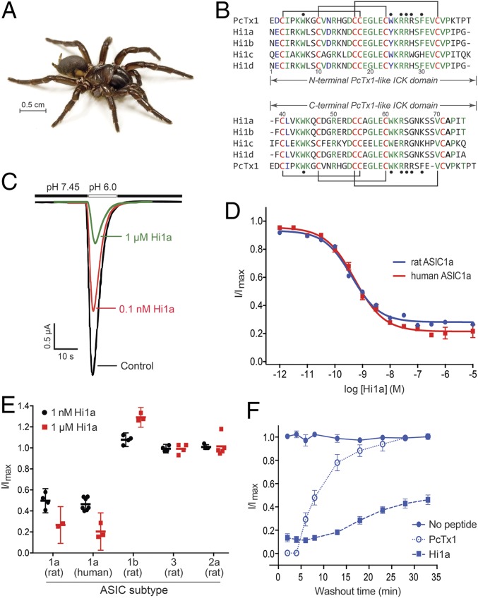
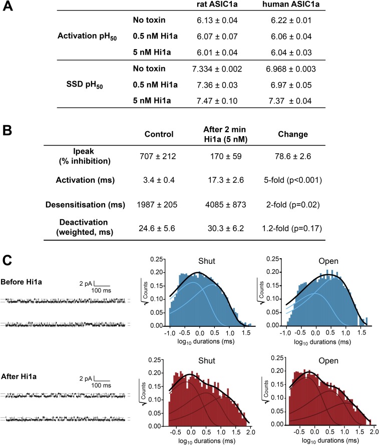
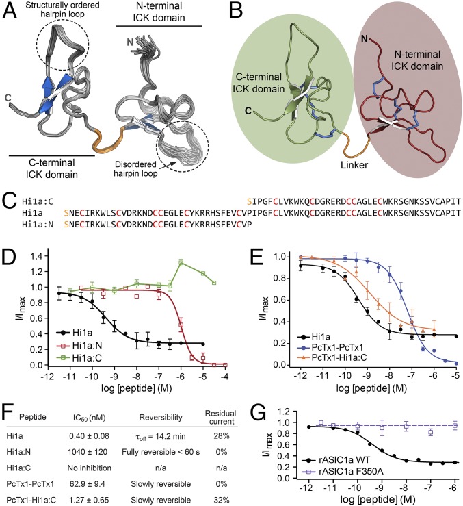
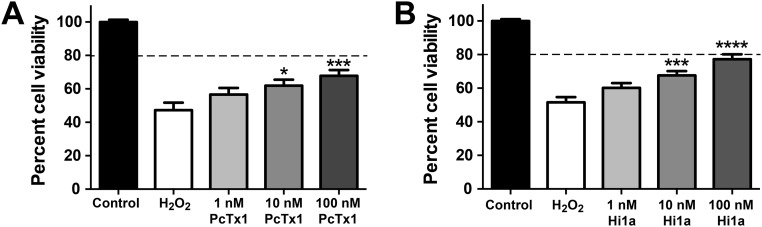
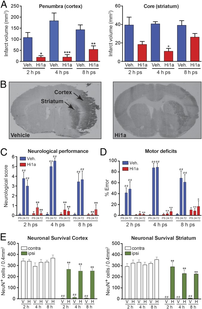
Stroke is the second-leading cause of death worldwide, yet there are no drugs available to protect the brain from stroke-induced neuronal injury. Acid-sensing ion channel 1a (ASIC1a) is the primary acid sensor in mammalian brain and a key mediator of acidosis-induced neuronal damage following cerebral ischemia. Genetic ablation and selective pharmacologic inhibition of ASIC1a reduces neuronal death following ischemic stroke in rodents. Here, we demonstrate that Hi1a, a disulfide-rich spider venom peptide, is highly neuroprotective in a focal model of ischemic stroke. Nuclear magnetic resonance structural studies reveal that Hi1a comprises two homologous inhibitor cystine knot domains separated by a short, structurally well-defined linker. In contrast with known ASIC1a inhibitors, Hi1a incompletely inhibits ASIC1a activation in a pH-independent and slowly reversible manner. Whole-cell, macropatch, and single-channel electrophysiological recordings indicate that Hi1a binds to and stabilizes the closed state of the channel, thereby impeding the transition into a conducting state. Intracerebroventricular administration to rats of a single small dose of Hi1a (2 ng/kg) up to 8 h after stroke induction by occlusion of the middle cerebral artery markedly reduced infarct size, and this correlated with improved neurological and motor function, as well as with preservation of neuronal architecture. Thus, Hi1a is a powerful pharmacological tool for probing the role of ASIC1a in acid-mediated neuronal injury and various neurological disorders, and a promising lead for the development of therapeutics to protect the brain from ischemic injury.

Keywords: acid-sensing ion channel 1a; ischemia; neuroprotection; stroke; venom peptide.


Conflict of interest statement

Conflict of interest statement: The authors' universities (The University of Queensland and Monash University) have jointly filed a patent application that covers use of the peptides described in this article (Hi1a–Hi1d).

Figures

Fig. 1.
Fig. 1.
Hi1a selectively inhibits ASIC1a. (A) Australian funnel-web spider H. infensa. Photo courtesy of Bastian Rast, ArachnoServer database (43). (B) Sequence alignment of PcTx1 with members of the Hi1a family. Identical and highly conserved residues are shown in green and blue, respectively, except for conserved cysteine residues, which are in red. The disulfide framework of PcTx1 is shown above and below the alignment. Black circles denote pharmacophore residues of PcTx1 (13, 20). (C) Representative current traces from Xenopus oocytes expressing rASIC1a in the absence (black) or presence (red and green) of Hi1a. Currents were evoked by a pH drop from 7.45 to 6.00. Note the incomplete current inhibition at a saturating concentration of Hi1a (1 μM). (D) Concentration-response curves for Hi1a inhibition of rASIC1a (blue) and hASIC1a (red). Fitting a Hill equation to the data yielded IC50 values of 0.40 ± 0.08 nM and 0.52 ± 0.06 nM, respectively. Data are mean ± SEM; n = 8. (E) Effect of 1 nM (black) and 1 μM (red) Hi1a on homomeric ASICs expressed in Xenopus oocytes. Hi1a has >2,000-fold selectivity for ASIC1a over these subtypes. Data are mean ± SEM; n = 5. (F) Recovery of rASIC1a currents following inhibition by Hi1a and PcTx1. Oocytes expressing rASIC1a were exposed to 10 nM PcTx1 or Hi1a at pH 7.45 for 120 s twice. Whole-cell currents were elicited by rapid switching from pH 7.45 to 6.00 every 60 s (and every 5 min following washout). Data are mean ± SEM; n = 5.
Fig. S1.
Fig. S1.
(A) SDS/PAGE gel illustrating various steps in the purification of recombinant Hi1a. Lanes are as follows: M, molecular mass markers; 1, insoluble material after lysis of Hi1a-expressing E. coli cells; 2, soluble extract resulting from cell lysis; 3, flow-through resulting from application of soluble cell extract to Ni-NTA beads; 4, eluate resulting from washing of Ni-NTA beads with 20 mM imidazole; 5, second 20 mM imidazole wash of Ni-NTA beads; 6, eluate resulting from washing of Ni-NTA beads with 400 mM imidazole, which is dominated by the MBP-Hi1a fusion protein; 7, same sample following cleavage of MBP-Hi1a fusion protein with TEV protease. (B, Bottom) Analytical RP-HPLC chromatogram showing pure recombinant Hi1a liberated by cleavage of the MBP-Hi1a fusion protein. (B, Top) Electrospray ionization mass spectrum of recombinant Hi1a (molecular weight 8,723 Da). (C) Table summarizing the calculated and observed monoisotopic (M+H+) masses for each of the recombinant peptides produced in this study.
Fig. S2.
Fig. S2.
Representative two-electrode voltage-clamp recordings showing the effects of PcTx1 and Hi1a on hASIC1a channels expressed in Xenopus oocytes. Whole-cell currents were elicited by rapidly switching the pH from 7.45 to 6.00 every 60 s (and every 5 min after washout). (A and B) Oocytes were exposed to 10 nM PcTx1 (A) or 10 nM rHi1a (B) at pH 7.45 for 120 s twice. (C) Recovery of hASIC1a currents during peptide washout compared with control oocytes (no peptide applied). The time constant for recovery from inhibition (τoff) was 0.99 min for PcTx1 and 31.8 min for Hi1a. Data are mean ± SEM. PcTx1, n = 3; Hi1a, n = 4.
Fig. 2.
Fig. 2.
Hi1a inhibits ASIC1a activation. (A) Effect of Hi1a on the pH-dependence of activation and SSD of rASIC1a (Left) and hASIC1a (Right). Activation curves were obtained by applying increasing concentrations of protons every 50 s. In the continued presence of protons (pH <∼7.2 for rASIC1a), ASICs rapidly desensitize and cannot reopen until sufficiently deprotonated (pH >∼7.3 for rASIC1a), a phenomenon known as SSD. SSD profiles were obtained by conditioning the channels for 120 s at decreasing pH. Data are mean ± SEM; n = 6. (B) Representative macropatch recordings from HEK293 cells expressing hASIC1a before (blue) and after (red) exposure to 5 nM Hi1a for 2 min. (Insert, Left) Expanded view of activation phase showing that Hi1a causes a marked reduction in the rate of current activation. (Insert, Right) Normalized deactivation phase currents showing that Hi1a has no significant effect on current deactivation. (C) Representative single-channel recording showing rapid activation of hASIC1a following a pH drop from 7.45 to 6.00 (Left, Middle). The lag time before channel activation is markedly increased in the presence of 5 nM Hi1a (Left, Bottom). (Right) Distribution of activation lag times before and after exposure to Hi1a (n = 10). *P < 0.001. (D, Left) Representative single-channel recordings before (Top) and after (Bottom) application of 5 nM Hi1a. (D, Right) Corresponding all-points amplitude histograms showing that Hi1a has no effect on hASIC1a current amplitude (∼1.0 pA).
Fig. S3.
Fig. S3.
(A) Table summarizing the effect of Hi1a on the activation and SSD profiles of rASIC1a and hASIC1a expressed in Xenopus oocytes. (B) Table summarizing kinetic parameters from macropatch experiments examining Hi1a-induced inhibition of hASIC1a expressed in HEK293 cells. (C) Analysis of the distributions of shut and open dwell times of single hASIC1a activity in HEK293 cells. Single-channel recordings (Left) obtained before preincubation with Hi1a (blue) revealed two shut components (Middle) and two open components (Right). Exposure of channels to Hi1a (red) led to observation of new components for both shut and open times. Notably, the new shut component (∼10 ms) contributed ∼40% of the shut distribution, whereas the new open component (∼8 ms) contributed only ∼13% of the total open times.
Fig. 3.
Fig. 3.
Hi1a is a double-knot peptide, with unique activity requiring both knots. (A) Solution structure of Hi1a (ensemble of 20 structures; PDB ID code 2N8F). A structured linker (orange) separates two closely apposed ICK domains. The β-hairpin loop in each ICK domain is highlighted. (B) Schematic of top-ranked structure from the Hi1a ensemble highlighting the N- and C-terminal ICK domains (red and green), linker (orange), and six disulfide bridges (blue). (C) Sequence alignment of recombinant Hi1a and recombinant Hi1a:N and Hi1a:C domains. The N-terminal serine residue (orange) is a vestige of the fusion protein cleavage site. (D) Concentration-response curves showing the effects of full-length Hi1a and Hi1a:N and Hi1a:C domains on rASIC1a. Hi1a:N fully inhibited rASIC1a, but with low potency (IC50 >1 μM), whereas Hi1a:C did not inhibit rASIC1a. Data are mean ± SEM; n = 6. (E) Effect of engineered double-knot peptides on rASIC1a. A peptide composed of two linked copies of PcTx1 fully inhibited rASIC1a with moderate potency (IC50 62.9 ± 9.4 nM; blue). In contrast, a chimeric double-knot peptide composed of an N-terminal PcTx1 domain joined to the C-terminal ICK domain of Hi1a (PcTx1-Hi1a:C) inhibited rASIC1a with similar potency as wild-type Hi1a (IC50 = 1.27 ± 0.65 nM; orange), and also caused incomplete inhibition at saturating concentrations. Data are mean ± SEM. PcTx1-PcTx1, n = 11; PcTx1-Hi1a:C, n = 6. (F) Pharmacologic properties of each peptide when tested against rASIC1a. Residual current is the percentage of pH-induced current remaining at a saturating concentration of peptide. Data are mean ± SEM; n = 6. (G) Concentration-dependent effects of Hi1a on wild-type and mutant (F350A) rASIC1a expressed in Xenopus oocytes.
Fig. S4.
Fig. S4.
Neuroprotective effects of PcTx1 (A) and Hi1a (B) following H2O2-induced oxidative injury in primary cortical neuron/astrocyte cultures. Cells were treated with 0.3 mM H2O2 in the absence and presence of Hi1a and PcTx1. Cell viability values were normalized, with those in the control group set to 100%. The horizontal dashed lines correspond to 80% cell viability. Data (mean ± SEM; n = 4) were analyzed using one-way ANOVA corrected for Dunnett’s multiple comparison test. *P < 0.05; ***P < 0.001; ****P < 0.0001 vs. H2O2. The neuroprotective effect of Hi1a was greater than that of PcTx1 (P < 0.05, two-way ANOVA). Primary mixed cortical neuronal-astrocytic cultures were prepared from embryonic day 18 Sprague–Dawley rats. Time-mated pregnant rats were euthanized with isoflurane, and cerebral cortices were dissected out in Earl’s Balanced Salt Solution (Life Technologies). Cortices were chemically digested with 1 mg/mL trypsin (Sigma-Aldrich) for 10 min, followed by 1 mg/mL trypsin inhibitor (Sigma-Aldrich) for 10 min. Tissues were washed and then triturated in DMEM/F12 Glutamax medium (Life Technologies). Cells were plated on poly-l-lysine–coated 48-well plates at a density of 100,000 cells/well in DMEM/F12 medium supplemented with 10% horse serum (Life Technologies), N-2 supplements (Life Technologies), 0.02 M d-glucose (Life Technologies), 2 mM sodium pyruvate (Life Technologies), 25 mM Hepes (Life Technologies), and 2 mM penicillin-streptomycin (Life Technologies). After 24 h, the medium was changed to fresh DMEM/F12. Primary cultures were used after being maintained for 7 d at 37 °C in a humidified 5% CO2 atmosphere incubator. Oxidative stress was induced by incubation with 0.3 mM H2O2 (Sigma Aldrich) prepared in DMEM/F12 in the presence and absence of Hi1a or PcTx1 (1, 10, or 100 nM) for 1 h. Medium containing H2O2 and ASIC1a inhibitors was removed and replaced with fresh DMEM/F12 medium containing different concentrations of inhibitors for 24 h at 37 °C. Cell viability was assessed by the 3-(4,5-dimethylthiazol-2-yl)-2,5-diphenyltetrazolium bromide (MTT) assay (Sigma Aldrich). In brief, 5 mg/mL of MTT was incubated with cell medium for 30 min at 37 °C to allow cells to cleave MTT and form a colored formazan product. The reaction was terminated by removing the medium containing MTT and replacing it with 100% DMSO for 10 min at room temperature. The formazan product was monitored at 570 nm with background subtraction at 630 nm.
Fig. 4.
Fig. 4.
Hi1a is highly neuroprotective in a realistic model of human stroke. (A) Infarct volumes in penumbral (cortical) and core (striatal) regions of damage following MCAO in conscious rats. Rats were administered i.c.v. vehicle (saline, blue) or Hi1a (2 ng/kg, red) at 2, 4, or 8 h poststroke (ps). Vehicle: 2 h, n = 10; 4 h, n = 7; 8 h, n = 9. Hi1a: 2 h, n = 5; 4 h, n = 7; 8 h, n = 10. Volumes were measured at 72 h poststroke and corrected for edema. *P < 0.05, **P < 0.01, ***P < 0.001 vs. vehicle (one-way ANOVA). (B) Coronal sections showing typical infarcted (darker area) and noninfarcted regions from rats treated with either vehicle or Hi1a (2 ng/kg) at 8 h after stroke. (C) Neurologic scores measured prestroke (PS) and at 24–72 h poststroke (ps). ##P < 0.01 vs. prestroke performance; **P < 0.01 vs. corresponding time in vehicle-treated group (two-way repeated-measures ANOVA followed by Tukey post hoc tests). (D) Motor score (% error in ledged beam test) measured prestroke and at 24–72 h poststroke. ##P < 0.01 vs. prestroke performance; *P < 0.05, **P < 0.01 vs. corresponding time in vehicle treated group (two-way repeated-measures ANOVA followed by Tukey post hoc tests). (E) Neuronal survival in cortical (Left) and striatal (Right) regions measured at 72 h poststroke. Data are expressed as number of NeuN-immunopositive (NeuN+) cells per 0.4 mm2 within occluded (ipsilateral) and nonoccluded (contralateral) hemispheres. **P < 0.01 vs. vehicle-treated group (ipsilateral side); ##P < 0.01 vs. matched region on noninfarcted hemisphere (two-way ANOVA followed by Tukey post hoc tests). All data are mean ± SEM.
Fig. S5.
Fig. S5.
(A) Effect of Hi1a on ET-1–induced cerebral vascular constriction. During the equilibration period, middle cerebral arteries generated spontaneous tone (46 ± 6% of maximum diameter; n = 7 arteries). Baseline diameters before the start of experiments were 65 ± 6 µm in the saline group (n = 3) and 55 ± 4 µm in the Hi1a group (n = 4). ET-1 evoked a similar degree of vasoconstriction in each group (27 ± 4% and 25 ± 1% in the saline and Hi1a groups, respectively). Under these conditions, Hi1a (1–100 nM) did not modify ET-1–evoked vasoconstriction, with vascular tone similar to that seen in time control experiments (saline/vehicle group). (B) The lack of effect of Hi1a was not due to impaired vessel integrity, because the arteries dilated fully to the vasodilator papaverine.

References

    1. Moskowitz MA, Lo EH, Iadecola C. The science of stroke: Mechanisms in search of treatments. Neuron. 2010;67(2):181–198. - PMC - PubMed
    1. Woodruff TM, et al. Pathophysiology, treatment, and animal and cellular models of human ischemic stroke. Mol Neurodegener. 2011;6(1):11. - PMC - PubMed
    1. Wey HY, Desai VR, Duong TQ. A review of current imaging methods used in stroke research. Neurol Res. 2013;35(10):1092–1102. - PMC - PubMed
    1. Choi DW, Rothman SM. The role of glutamate neurotoxicity in hypoxic-ischemic neuronal death. Annu Rev Neurosci. 1990;13:171–182. - PubMed
    1. Liu R, Yuan H, Yuan F, Yang SH. Neuroprotection targeting ischemic penumbra and beyond for the treatment of ischemic stroke. Neurol Res. 2012;34(4):331–337. - PubMed

Publication types

MeSH terms

Associated data