. 2017 Apr 18;114(16):E3334-E3343.
doi: 10.1073/pnas.1616467114. Epub 2017 Mar 20.

Neuroendocrine androgen action is a key extraovarian mediator in the development of polycystic ovary syndrome

Affiliations

Neuroendocrine androgen action is a key extraovarian mediator in the development of polycystic ovary syndrome

Aimee S L Caldwell et al. Proc Natl Acad Sci U S A. .

Abstract

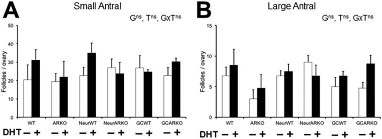
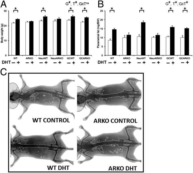
Polycystic ovary syndrome (PCOS) is a complex hormonal disorder characterized by reproductive, endocrine, and metabolic abnormalities. As the origins of PCOS remain unknown, mechanism-based treatments are not feasible and current management relies on treatment of symptoms. Hyperandrogenism is the most consistent PCOS characteristic; however, it is unclear whether androgen excess, which is treatable, is a cause or a consequence of PCOS. As androgens mediate their actions via the androgen receptor (AR), we combined a mouse model of dihydrotestosterone (DHT)-induced PCOS with global and cell-specific AR-resistant (ARKO) mice to investigate the locus of androgen actions that mediate the development of the PCOS phenotype. Global loss of the AR reveals that AR signaling is required for all DHT-induced features of PCOS. Neuron-specific AR signaling was required for the development of dysfunctional ovulation, classic polycystic ovaries, reduced large antral follicle health, and several metabolic traits including obesity and dyslipidemia. In addition, ovariectomized ARKO hosts with wild-type ovary transplants displayed normal estrous cycles and corpora lutea, despite DHT treatment, implying extraovarian and not intraovarian AR actions are key loci of androgen action in generating the PCOS phenotype. These findings provide strong evidence that neuroendocrine genomic AR signaling is an important extraovarian mediator in the development of PCOS traits. Thus, targeting AR-driven mechanisms that initiate PCOS is a promising strategy for the development of novel treatments for PCOS.

Keywords: PCOS; androgen; animal model; neuroendocrine.


Conflict of interest statement

The authors declare no conflict of interest.

Figures

Fig. 1.
Fig. 1.
Experimental design. For this study, PCOS was induced in wild-type, global, neuron-specific, and granulosa cell-specific androgen receptor knockout mice by s.c. inserting dihydrotestosterone implants in the mice for 3 mo. Control mice were implanted with blank implants. Body weight, estrous cycling, blood pressure, fasting glucose, oral glucose tolerance, and insulin tolerance were assessed before collection of serum and tissues at 16 wk of age.
Fig. 2.
Fig. 2.
Neuron but not granulosa cell AR signaling is required for the development of anovulation in the PCOS mouse model. Loss of extraovarian AR signaling ameliorates the development of acyclicity and anovulation in the PCOS mouse model. (A) Average number of full estrous cycles completed in a 2-wk period, confirming DHT-induced acyclicity in WT mice and showing no development of acyclicity in DHT-induced PCOS ARKO female mice. Data are the mean ± SEM; n = 5 to 9 per genotype/treatment group. (B) Average number of corpora lutea per ovary, showing no development of anovulation in DHT-induced PCOS ARKO or NeurARKO female mice. Data are the mean ± SEM; n = 3 to 5 per genotype/treatment group. (C) Histological sections of representative ovaries from each treatment group, showing the maintenance of ovulations (indicated by the presence of corpora lutea) in DHT-induced PCOS ARKO and NeurARKO ovaries. (D) Percentage of cycling females in a 2-wk period, showing no development of acyclicity in DHT-induced PCOS ARKO ovariectomized females transplanted with WT ovaries. (E) Histological sections of representative ovaries from each treatment group, showing corpora lutea in DHT-induced PCOS ARKO ovariectomized females transplanted with WT ovaries, indicative of recent ovulation. G, genotype; ns, no significant difference; T, DHT treatment; *, significant difference; star, corpora lutea; triangle, arrested antral follicle. (Magnification: 10×.) (*P < 0.05, two-way ANOVA.)
Fig. S1.
Fig. S1.
Characterization of estrous-cycle patterns. Estrous-cycle pattern in representative females. D, diestrus; E, estrus; M, metestrus; P, proestrus.
Fig. 3.
Fig. 3.
Global and neuron but not granulosa cell loss of AR signaling protects against the development of the classic polycystic ovarian phenotype in the PCOS mouse model. (A) Ovary weights, confirming DHT-induced reduction in ovarian weight in WT mice, and showing no reduction in ovarian weight in DHT-induced PCOS ARKO female mice. Data are the mean ± SEM; n = 5 to 9 per genotype/treatment group. (B) Average number of atretic cyst-like follicles per ovary, displaying no significant development of cysts in DHT-induced PCOS ARKO females. Data are the mean ± SEM; n = 4 ovaries per genotype/treatment group. (C) Histological sections of representative ovaries, showing development of the classic PCOS ovarian phenotype in DHT-induced PCOS WT but not ARKO and NeurARKO ovaries. (D) Histological section of a representative cyst found in all groups treated with DHT. Cysts display a thinned and discordant granulosa cell layer, a dispersed theca cell layer (arrow), and an oocyte with a fragmented nucleolus that has lost connection with most or all of its surrounding granulosa cells. G, genotype; ns, no significant difference; T, DHT treatment; *, significant difference. (Magnification: 10×.) (*P < 0.05, two-way ANOVA.)
Fig. 4.
Fig. 4.
Global and neuron but not granulosa cell loss of AR signaling protects against the reduction in large antral follicle health in the PCOS mouse model. (A) Percentage of unhealthy small antral follicles per ovary, confirming DHT-induced increase in unhealthy small antral follicles in WT mice and showing no significant decrease in small antral follicle health in DHT-induced PCOS ARKO ovaries. Data are the mean ± SEM; n = 4 ovaries per genotype/treatment group. (B) Percentage of unhealthy large antral follicles per ovary, showing no significant decrease in large antral follicle health in DHT-induced PCOS ARKO or NeurARKO female mice. Data are the mean ± SEM; n = 4 ovaries per genotype/treatment group. (C) Average thickness of granulosa cell layer per follicle, displaying no reduction in granulosa cell-layer thickness in DHT-induced PCOS ARKO or GCARKO ovaries. Data are the mean ± SEM; n = 7 to 24 follicles per genotype/treatment group. (D) Average percentage of theca area per follicle, showing no significant increase in theca cell area in DHT-induced PCOS ARKO ovaries. Data are the mean ± SEM; n = 7 to 24 follicles per genotype/treatment group. (E) Histological sections representing the effects of DHT on granulosa cell-layer thickness (ii, double-ended arrow) and percentage of theca area (iv, white line) compared with controls (i and iii, respectively). G, genotype; ns, no significant difference; T, DHT treatment; *, significant difference. (Magnification: 10×.) (*P < 0.05, two-way ANOVA.)
Fig. S2.
Fig. S2.
Quantification of antral follicle populations. Average number of small antral (A) and large antral (B) follicles per ovary. Data are the mean ± SEM; n = 4 ovaries per genotype/treatment group.
Fig. 5.
Fig. 5.
Global and neuron but not granulosa cell loss of AR signaling protects against the development of obesity in the PCOS mouse model. (A) Body weight, confirming DHT-induced increased body weight in WT mice and showing no significant increase in body weight in DHT-induced PCOS ARKO or NeurARKO female mice. Data are the mean ± SEM; n = 5 to 9 mice per genotype/treatment group. (B) Parametrial fat deposit weight, showing no significant increase in adiposity in DHT-induced PCOS ARKO or NeurARKO female mice. Data are the mean ± SEM; n = 5 to 9 per genotype/treatment group. (C) Representative dual-energy X-ray absorptiometry images, showing no development of obesity in DHT-induced PCOS ARKO female mice. G, genotype; ns, no significant difference; T, DHT treatment; *, significant difference. (*P < 0.05, two-way ANOVA.)
Fig. 6.
Fig. 6.
Global loss of AR signaling protects against the reduction in adiponectin levels, adipocyte hypertrophy, and altered glucose homeostasis in the PCOS mouse model. (A) Serum levels of adiponectin, confirming DHT-induced suppression of serum adiponectin levels in WT mice and showing no significant reduction in adiponectin levels in DHT-induced PCOS ARKO female mice. Data are the mean ± SEM; n = 5 to 7 per genotype/treatment group. (B) Adipocyte size, showing no development of adipocyte hypertrophy in DHT-induced PCOS ARKO female mice. Data are the mean ± SEM; n = 3 sections per mouse, 3 mice per genotype/treatment group. (C) Histological sections of representative parametrial fat pads from each treatment group, showing no development of adipocyte hypertrophy in DHT-induced PCOS ARKO female mice. (D) Average fasting glucose levels showing no significant increase in glucose levels in DHT-induced PCOS ARKO female mice. Data are the mean ± SEM; n = 5 to 9 per genotype/treatment group. (E) Area under the curve (AUC) analysis of the oral glucose tolerance test, showing an overall effect of DHT treatment. Data are the mean ± SEM; n = 5 to 9 per genotype/treatment group. G, genotype; ns, no significant difference; T, DHT treatment; *, significant difference. (Magnification: 40×.) (*P < 0.05, two-way ANOVA.)
Fig. S3.
Fig. S3.
Characterization of insulin tolerance. AUC analysis of insulin tolerance, showing an effect of genotype only (*P < 0.01, two-way ANOVA). Data are the mean ± SEM; n = 5 to 9 per genotype/treatment group.
Fig. 7.
Fig. 7.
Global and neuron but not granulosa cell loss of AR signaling ameliorates the development of dyslipidemia in the PCOS mouse model. (A) Serum cholesterol levels, confirming DHT-induced increased serum cholesterol in WT mice and showing no significant increase in cholesterol levels in DHT-induced PCOS ARKO or NeurARKO female mice. Data are the mean ± SEM; n = 5 to 7 per genotype/treatment group. (B) Serum triglyceride levels, showing an overall effect of DHT treatment and a nonsignificant trend to increased triglyceride levels in all DHT-treated groups, apart from DHT-induced PCOS ARKO and NeurARKO female mice. Data are the mean ± SEM; n = 5 to 7 per genotype/treatment group. (C) Systolic blood pressure, displaying no significant difference between control and DHT-treated PCOS mice of any genotype. Data are the mean ± SEM; n = 5 to 9 per genotype/treatment group. (D) Analysis of liver steatosis by oil red O staining, showing no significant increase in the presence of steatosis in DHT-induced PCOS ARKO female mice. Data are the mean ± SEM; n = 3 sections per mouse, 3 mice per genotype/treatment group. (D) Histological sections of representative liver sections stained with oil red O, showing reduced lipid staining in DHT-induced PCOS ARKO livers. G, genotype; ns, no significant difference; T, DHT treatment; *, significant difference. (Magnification: 40×.) (*P < 0.05, two-way ANOVA.)
Fig. S4.
Fig. S4.
Characterization of global and tissue-specific ARKO mice. Representation of RT-PCR androgen receptor analyses using cDNA extracted from WT, ARKO, NeurWT, NeurARKO, GCWT, and GCARKO ovary, uterus, brain, and pituitary. The intact AR exon 3 PCR product is 288 bp long, whereas the excised AR exon 3 PCR product is 171 bp. Mouse β-actin was used as an internal control, with a PCR product of 431 bp.

Comment in

References

    1. Fauser BC, et al. Consensus on women’s health aspects of polycystic ovary syndrome (PCOS): The Amsterdam ESHRE/ASRM-Sponsored 3rd PCOS Consensus Workshop Group. Fertil Steril. 2012;97(1):28–38.e25. - PubMed
    1. Gorry A, White DM, Franks S. Infertility in polycystic ovary syndrome: Focus on low-dose gonadotropin treatment. Endocrine. 2006;30(1):27–33. - PubMed
    1. Dumesic DA, et al. Scientific statement on the diagnostic criteria, epidemiology, pathophysiology, and molecular genetics of polycystic ovary syndrome. Endocr Rev. 2015;36(5):487–525. - PMC - PubMed
    1. Shorakae S, Boyle J, Teede H. Polycystic ovary syndrome: A common hormonal condition with major metabolic sequelae that physicians should know about. Intern Med J. 2014;44(8):720–726. - PubMed
    1. Nisenblat V, Norman RJ. Androgens and polycystic ovary syndrome. Curr Opin Endocrinol Diabetes Obes. 2009;16(3):224–231. - PubMed

Publication types

MeSH terms