. 2017:2017:6543237.
doi: 10.1155/2017/6543237. Epub 2017 Feb 22.

The Antimalarial Chloroquine Suppresses LPS-Induced NLRP3 Inflammasome Activation and Confers Protection against Murine Endotoxic Shock

Affiliations

The Antimalarial Chloroquine Suppresses LPS-Induced NLRP3 Inflammasome Activation and Confers Protection against Murine Endotoxic Shock

Xiaoli Chen et al. Mediators Inflamm. 2017.

Abstract

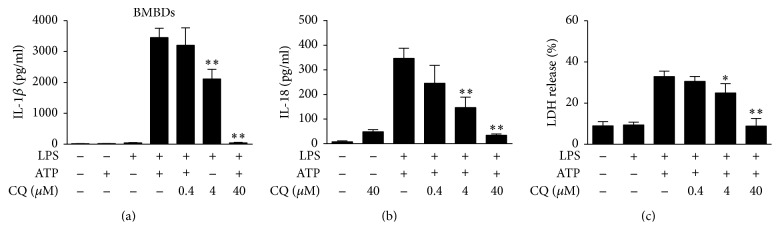
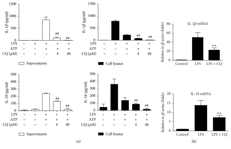
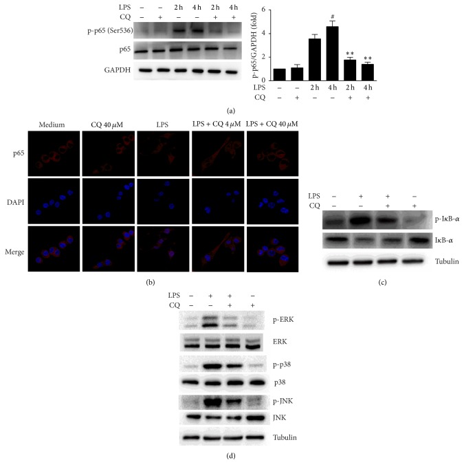
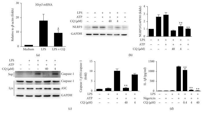
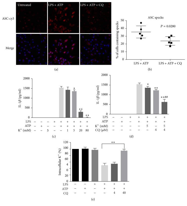
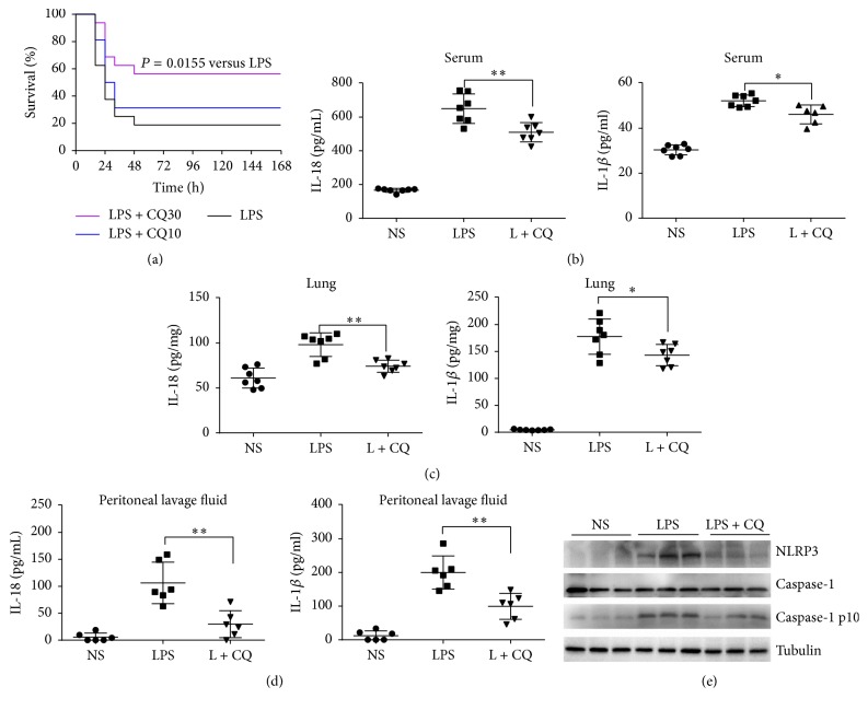
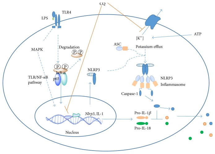
Activation of the NLRP3 inflammasome, which catalyzes maturation of proinflammatory cytokines like IL-1β and IL-18, is implicated and essentially involved in many kinds of inflammatory disorders. Chloroquine (CQ) is a traditional antimalarial drug and also possesses an anti-inflammatory property. In this study, we investigated whether CQ suppresses NLRP3 inflammasome activation and thereby confers protection against murine endotoxic shock. CQ attenuated NF-κB and MAPK activation and prohibited expression of IL-1β, IL-18, and Nlrp3 in LPS treated murine bone marrow-derived macrophages (BMDMs), demonstrating its inhibitory effect on the priming signal of NLRP3 activation. Then, CQ was shown to inhibit caspase-1 activation and ASC specks formation in BMDMs, which indicates that CQ also suppresses inflammasome assembly, the second signal for NLRP3 inflammasome activation. In a murine endotoxic shock model, CQ effectively improved survival and markedly reduced IL-1β and IL-18 production in serum, peritoneal fluid, and lung tissues. Moreover, CQ reduced protein levels of NLRP3 and caspases-1 p10 in lung homogenates of mice with endotoxic shock, which may possibly explain its anti-inflammatory activity and life protection efficacy in vivo. Overall, our results demonstrate a new role of CQ that facilitates negative regulation on NLRP3 inflammasome, which thereby confers protection against lethal endotoxic shock.


Conflict of interest statement

The authors declare that there are no competing interests regarding the publication of this paper.

Figures

Figure 1
Figure 1
CQ suppress IL-1β and IL-18 secretion in LPS- and ATP-primed BMDMs. BMDMs were pretreated with or without 0.4, 4, and 40 μM CQ for 1 h and then stimulated with LPS (100 ng/ml) for 4 h and ATP (2 mM) for an additional 1 h. The IL-1β and IL-18 levels in culture supernatants were determined by ELISA (n = 3). ∗∗P < 0.01 versus LPS plus ATP group (a-b). LDH release from BMDMs was measured by LDH assay (c). P < 0.05, ∗∗P < 0.01 versus LPS plus ATP group. All data were expressed as mean ± SEM in triplicate and are representative of three independent experiments.
Figure 2
Figure 2
CQ inhibits transcription and maturation of IL-1β and IL-18. (a) BMDMs were pretreated with CQ (4, 40 μM) for 1 h, primed with LPS (100 ng/ml) for 4 h, and stimulated with ATP (2 mM) for an additional 1 h. Cell lysates and supernatants were prepared and IL-1β level was detected by ELISA. ∗∗P < 0.01 versus LPS plus ATP group. (b) IL-1β and IL-18 mRNA expressions were measured by qRT-PCR. BMDMs were pretreated with CQ (40 μM) for 1 h and then primed with LPS (100 ng/ml) for 2 h. Data are shown as the means ± SEM (n = 3). ∗∗P < 0.01 versus LPS group; ##P < 0.01 versus LPS plus ATP group.
Figure 3
Figure 3
CQ restrains NF-κB p65 phosphorylation and nuclear localization. (a) Western blot of phospho-NF-κB p65 (Ser536). Cells were pretreated with 40 μM CQ for 1 h and then 100 ng/ml LPS treated for 2 h and 4 h, respectively. Total cell protein was collected for western blot of NF-κB p65 phosphorylation and GAPDH. #P < 0.05 versus LPS group (2 h). ∗∗P < 0.01 versus LPS group (4 h). (b) Cells were pretreated with 40 μM CQ for 1 h and then stimulated by LPS for 30 min. IκB-α and phospho-IκB-α levels were detected by western blot. (c) Confocal images of nuclear translocation of NF-κB p65. Cells were pretreated with different concentrations of CQ for 1 h, followed by 100 ng/ml LPS treatment for 2 h. Cells were stained by anti-NF-κB p65 (red) antibodies and DAPI (blue) for nuclear staining. (d) Cells were treated as in (b). ERK, p38, and JNK and their phosphorylated forms (p-IκB-α, p-ERK, p-p38, and p-JNK) in cell lysates were detected by western blot.
Figure 4
Figure 4
CQ dampens NLRP3 inflammasome activation. BMDMs were pretreated with CQ (4, 40 μM) for 1 h and then stimulated with LPS (100 ng/ml) for 4 h and ATP (2 mM) for an additional 1 h. (a) Nlrp3 mRNA expression was measured by qRT-PCR. (b) NLRP3 and GAPDH were measured in cell lysates by western blot. (c) Western blot analysis of cleaved caspase-1 (p10) in culture supernatants (Sup) or of procaspase-1, ASC, and tubulin in cell lysates (Lys). (d) BMDMs were pretreated with CQ at the indicated concentration for 1 h and then stimulated with LPS (100 ng/ml) for 4 h and nigericin (10 μM) for 30 min. Production of mature IL-1β in supernatants was measured by ELISA. Data are shown as means ± SEM; P < 0.05, ∗∗P < 0.01 versus LPS plus ATP group.
Figure 5
Figure 5
CQ restrains ASC speck formation and potassium efflux. (a) Representative immunofluorescence images of ASC speck formation in LPS-primed BMDMs stimulated with ATP in the presence or absence of CQ (20 μM) were acquired by confocal microscopy. (b) A percentage of cells containing specks were quantified in at least five distinct fields. (c, d) LPS-primed BMDMs were treated for 1 h with 2 mM ATP in a medium containing the specified [K+], and the secreted IL-1β was measured. (e) CQ pretreatment BMDMs were treated with LPS plus ATP, and the intracellular content of K+ was detected. Data are shown as means ± SEM; ##P < 0.01 versus CQ pretreatment (4 μM) group; P < 0.05, ∗∗P < 0.01 versus LPS plus ATP group.
Figure 6
Figure 6
CQ protects mice against lethal endotoxic shock by suppressing NLRP3 inflammasome activation. CQ improved survival in endotoxic shock murine model. BALB/c murine mice (n = 16 per group) were injected with a lethal dose of LPS (15 mg/kg) alone or together with CQ (10, 30 mg/kg). (a) Survival in each group was observed for 168 h and the log-rank test was used for intergroup comparison. P < 0.05 versus LPS group. (b) IL-18 and IL-1β levels in serum at 6 h were measured by ELISA. (c) IL-18 and IL-1β levels in lung homogenates at 6 h were measured by ELISA (n = 7 per group). (d) IL-18 and IL-1β levels in peritoneal lavage fluid at 6 h were measured by ELISA (n = 6 per group). Data are shown as means ± SEM. P < 0.05, ∗∗P < 0.01 versus LPS group. (e) Caspase-1 activation and NLRP3 expression in the lung were detected by western blot (n = 3).
Figure 7
Figure 7
The proposed mechanism of CQ for the inhibition of NLRP3 inflammasome activation.

References

    1. Lamkanfi M., Dixit V. M. Inflammasomes and their roles in health and disease. Annual Review of Cell and Developmental Biology. 2012;28:137–161. doi: 10.1146/annurev-cellbio-101011-155745. - DOI - PubMed
    1. Grellier N., Deray G., Yousfi A., Khodari W., Bouaita R., Belkacemi Y. Carence martiale fonctionnelle, inflammation et fatigue après radiothérapie. Bulletin Du Cancer. 2015;102(9):780–785. - PubMed
    1. Dinarello C. A. An expanding role for interleukin-1 blockade from gout to cancer. Molecular Medicine. 2014;20(supplement 1):S43–S58. doi: 10.2119/molmed.2014.00232. - DOI - PMC - PubMed
    1. Ballak D. B., Stienstra R., Tack C. J., Dinarello C. A., van Diepen J. A. IL-1 family members in the pathogenesis and treatment of metabolic disease: focus on adipose tissue inflammation and insulin resistance. Cytokine. 2015;75(2):280–290. doi: 10.1016/j.cyto.2015.05.005. - DOI - PMC - PubMed
    1. Palomo J., Dietrich D., Martin P., Palmer G., Gabay C. The interleukin (IL)-1 cytokine family—balance between agonists and antagonists in inflammatory diseases. Cytokine. 2015;76(1):25–37. doi: 10.1016/j.cyto.2015.06.017. - DOI - PubMed

MeSH terms

LinkOut - more resources