. 2018 Mar;38(2):421-430.
doi: 10.1007/s10571-017-0485-1. Epub 2017 Mar 20.

Suppression of the Smurf1 Expression Inhibits Tumor Progression in Gliomas

Affiliations

Suppression of the Smurf1 Expression Inhibits Tumor Progression in Gliomas

Hao Chang et al. Cell Mol Neurobiol. 2018 Mar.

Abstract

Glioblastoma, one of the common malignant brain tumors, results in the highly death, but its underlying molecular mechanisms remain unclear. Smurf1, a member of Nedd4 family of HECT-type ligases, has been reported to contribute to tumorigenicity through several important biological pathways. Recently, it was also found to participate in modulate cellular processes, including morphogenesis, autophagy, growth, and cell migration. In this research, we reported the clinical guiding significance of the expression of Smurf1 in human glioma tissues and cell lines. Western blotting analysis discovered that the expression of Smurf1 was increased with WHO grade. Immunohistochemistry levels discovered that high expression of Smurf1 is closely consistent with poor prognosis of glioma. In addition, suppression of Smurf1 can reduce cell invasion and increase the E-cadherin expression, which is a marker of invasion. Our study firstly demonstrated that Smurf1 may promote glioma cell invasion and suppression of the Smurf1 may provide a novel treatment strategy for glioma.

Keywords: Apoptosis; Glioma; Migration; Smurf1.


Conflict of interest statement

The authors declare that they have no conflict of interest.

Figures

Fig. 1
Fig. 1
The Smurf1 expression in human normal tissues, glioma tissues, and cells. a, c Smurf1 expression in glioma tissues (grades II–IV), normal brain tissues, normal human gliocyte HEB cells, U87MG, U251MG, U118, and A172 glioma cells was analyzed by Western blot analysis. GAPDH was used as a control for protein load and integrity. b, d The bar chart shows the ratio of Smurf1 protein to GAPDH by quantitative analysis. The data are mean ± SD of three independent experiments
Fig. 2
Fig. 2
The correlation between Smurf1 and the grade of glioma, E-cadherin, and glioma outcome. a Paraffin-embedded glioma tissue sections (including grades IIIV) were stained with anti-Smurf1 antibodies and anti-E-cadherin antibodies, and counterstained with hematoxylin (SP × 400). b The relationship between Smurf1 and E-cadherin. c Kaplan–Meier survival curve for patterns of patients with glioma and the expression of Smurf1 in 98 patients with glioma. Patients in the high expression Smurf1 group had an apparently shorter overall survival (P < 0.01)
Fig. 3
Fig. 3
Silencing of Smurf1 inhibits the migration of glioma cells. a Western blot analyzed Smurf1 expression after transfected with Smurf1-siRNA in U87-MG cells and results showed that siSmurf1-4 achieved the best knockout effect. b The bar chart shows the ratio of Smurf1 protein to GAPDH for the above by densitometry. The data are mean ± SD. (*P < 0.05 compared with the control). c, d U87-MG cells transfected with Smurf1 siRNA and si-NC were examined in wound-healing assays. Images were captured at 0 and 48 h from an inverted Leica phase-contrast microscope (200× magnification). *P < 0.05. c, d Wound-healing assays indicate the effect of Smurf1 expression on glioma cell migration ability. Each time point is collected from three independent experiments
Fig. 4
Fig. 4
Knockdown of Smurf1 inhibits the glioma cells invasion. a, b Transwell assays indicate the effect of Smurf1 expression on glioma cell migration ability. Each time point is collected from three independent experiments. c Western blot analysis of E-cadherin, vimentin in control, si-NC, and siSmurf1 cells
Fig. 5
Fig. 5
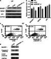
Effect of Smurf1 down-expression on cell apoptosis in glioma. a, b Three proteins, Smurf1, MDM2, and p53 in control, si-NC and si-Smurf1-4-transfected U87MG cells were analyzed by Western blot analysis. c U87MG cells were treated with si-NC and si-Smurf1-4, respectively, and then assayed by Becton–Dickinson flow cytometer BD FACScan according to the manufacturer’s instructions. Interference of Smurf1 significantly promoted apoptosis in U87MG cells. d Two apoptotic marker proteins, cleaved caspase-3 and cleaved PARP in si-NC- and si-Smurf1-4-transfected U87MG cells were analyzed by Western blot analysis

References

    1. Ahmed R, Oborski MJ, Hwang M, Lieberman FS, Mountz JM (2014) Malignant gliomas: current perspectives in diagnosis, treatment, and early response assessment using advanced quantitative imaging methods. Cancer Manag Res 6:149–170. doi:10.2147/CMAR.S54726 - PMC - PubMed
    1. Bai RY, Staedtke V, Riggins GJ (2011) Molecular targeting of glioblastoma: drug discovery and therapies. Trends Mol Med 17(6):301–312. doi:10.1016/j.molmed.2011.01.011 - PMC - PubMed
    1. Cao Y, Zhang L (2013) A Smurf1 tale: function and regulation of an ubiquitin ligase in multiple cellular networks. Cell Mol Life Sci 70(13):2305–2317. doi:10.1007/s00018-012-1170-7 - PMC - PubMed
    1. Chen J, Sun J, Yang L, Yan Y, Shi W, Shi J, Huang Q, Chen J, Lan Q (2015) Upregulation of B23 promotes tumor cell proliferation and predicts poor prognosis in glioma. Biochem Biophys Res Commun 466(1):124–130. doi:10.1016/j.bbrc.2015.08.118 - PubMed
    1. Cohen ZR, Ramishetti S, Peshes-Yaloz N, Goldsmith M, Wohl A, Zibly Z, Peer D (2015) Localized RNAi therapeutics of chemoresistant grade IV glioma using hyaluronan-grafted lipid-based nanoparticles. ACS Nano 9(2):1581–1591. doi:10.1021/nn506248s - PubMed

Substances

LinkOut - more resources