

Acute hyperventilation increases the central venous-to-arterial PCO2 difference in stable septic shock patients
- PMID: 28321801
- PMCID: PMC5359263
- DOI: 10.1186/s13613-017-0258-5
Acute hyperventilation increases the central venous-to-arterial PCO2 difference in stable septic shock patients
Abstract
Background: To evaluate the effects of acute hyperventilation on the central venous-to-arterial carbon dioxide tension difference (∆PCO2) in hemodynamically stable septic shock patients.
Methods: Eighteen mechanically ventilated septic shock patients were prospectively included in the study. We measured cardiac index (CI), ∆PCO2, oxygen consumption (VO2), central venous oxygen saturation (ScvO2), and blood gas parameters, before and 30 min after an increase in alveolar ventilation (increased respiratory rate by 10 breaths/min).
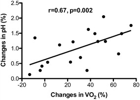
Results: Arterial pH increased significantly (from 7.35 ± 0.07 to 7.42 ± 0.09, p < 0.001) and arterial carbon dioxide tension decreased significantly (from 44.5 [41-48] to 34 [30-38] mmHg, p < 0.001) when respiratory rate was increased. A statistically significant increase in VO2 (from 93 [76-105] to 112 [95-134] mL/min/m2, p = 0.002) was observed in parallel with the increase in alveolar ventilation. While CI remained unchanged, acute hyperventilation led to a significant increase in ∆PCO2 (from 4.7 ± 1.0 to 7.0 ± 2.6 mmHg, p < 0.001) and a significant decrease in ScvO2 (from 73 ± 6 to 67 ± 8%, p < 0.001). A good correlation was found between changes in arterial pH and changes in VO2 (r = 0.67, p = 0.002). Interestingly, we found a strong association between the increase in VO2 and the increase in ∆PCO2 (r = 0.70, p = 0.001).
Conclusions: Acute hyperventilation provoked a significant increase in ∆PCO2, which was the result of a significant increase in VO2 induced by hyperventilation. The clinician should be aware of the effects of acute elevation of alveolar ventilation on ∆PCO2.
Keywords: Acute hyperventilation; Central venous oxygen saturation; Central venous-to-arterial CO2 tension gap; Oxygen consumption; Septic shock.
Figures

References
-
- Mallat J, Pepy F, Lemyze M, Gasan G, Vangrunderbeeck N, Tronchon L, et al. Central venous-to-arterial carbon dioxide partial pressure difference in early resuscitation from septic shock: a prospective observational study. Eur J Anaesthesiol. 2014;31(7):371–380. doi: 10.1097/EJA.0000000000000064. - DOI - PubMed
LinkOut - more resources
Full Text Sources
Other Literature Sources
Miscellaneous
