Review
. 2016;36(4):283-314.
doi: 10.1615/CritRevImmunol.2017019158.

The Goldilocks Conundrum: NLR Inflammasome Modulation of Gastrointestinal Inflammation during Inflammatory Bowel Disease

Affiliations
Review

The Goldilocks Conundrum: NLR Inflammasome Modulation of Gastrointestinal Inflammation during Inflammatory Bowel Disease

Veronica M Ringel-Scaia et al. Crit Rev Immunol. 2016.

Abstract

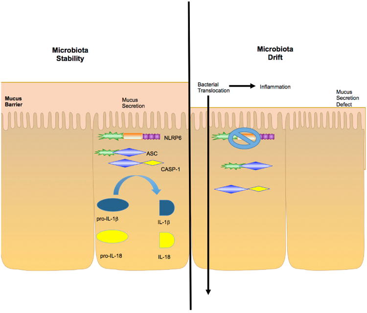
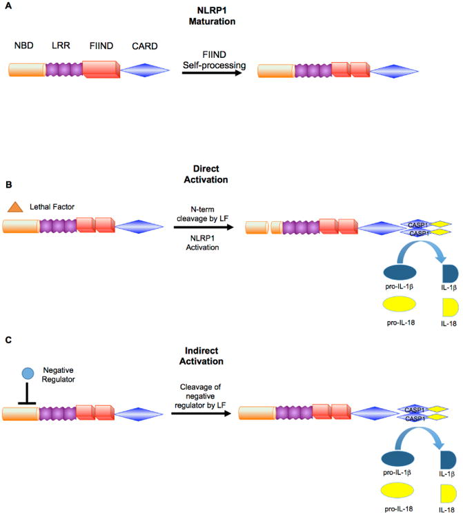
Recent advances have revealed significant insight into inflammatory bowel disease (IBD) pathobiology. Ulcerative colitis and Crohn's disease, the chronic relapsing clinical manifestations of IBD, are complex disorders with genetic and environmental influences. These diseases are associated with the dysregulation of immune tolerance, excessive inflammation, and damage to the epithelial cell barrier. Increasing evidence indicates that pattern recognition receptors, including Toll-like receptors (TLRs) and nucleotide-binding domain and leucine-rich repeat-containing proteins (NLRs), function to maintain immune system homeostasis, modulate the gastrointestinal microbiome, and promote proper intestinal epithelial cell regeneration and repair. New insights have revealed that NLR family members are essential components in maintaining this immune system homeostasis. To date, the vast majority of studies associated with NLRs have focused on family members that form a multiprotein signaling platform called the inflammasome. These signaling complexes are responsible for the cleavage and activation of the potent pleotropic cytokines IL-1β and IL-18, and they facilitate a unique form of cell death defined as pyroptosis. In this review, we summarize the current paradigms associated with NLR inflammasome maintenance of immune system homeostasis in the gastrointestinal system. New concepts related to canonical and noncanonical inflammasome signaling, as well as the implications of classical and alternative inflammasomes in IBD pathogenesis, are also reviewed.


Figures

Fig. 1
Fig. 1
Inflammatory Bowel Disease Pathogenesis. Crohn's disease (CD) and ulcerative colitis (UC) are complex disorders, which are driven by poorly understood genetic and environmental influences. These disorders are strongly associated with dysfunctional and overzealous immune system signaling in the gastrointestinal tract. It is also clear that dysbiosis and perhaps even mild-to-moderate changes in specific host microbiome populations can dramatically influence IBD progression and prognosis. Each of these mechanisms function in synergy to drive IBD pathobiology.
Fig. 2
Fig. 2
The Canonical and Noncanonical Inflammasome. The activation of the canonical NLR Inflammasome requires priming, typically via activation of a Toll-like receptor (TLR). Following ligation of the TLR, MyD88 is recruited and subsequently leads to the activation of NF-κB signaling. Activated NF-κB promotes the transcription of pro-IL-1β and pro-IL-18. Many mechanisms have been proposed for the activation of the NLR Inflammasome once priming has occurred: direct PAMP sensing, K+ efflux, pore forming channels and toxins, Ca2+ influx, reactive oxygen species from the mitochondria, mitochondrial DNA, cardiolipin translocation, FADD, caspase-8 and rupture of phagosomes. Upon activation from one of these mechanisms, the NLR, ASC and caspase-1 come together to form the core unit of the Inflammasome. This assembly leads to the cleavage of pro-IL-1β and pro-IL-18 into their mature forms and the induction of the pro-Inflammatory form of cell death defined as pyroptosis. In noncanonical Inflammasome activation, type I interferon is stimulated via the TLR-TRIF–mediated pathway, which drives STAT1 activation and leads to the induction of caspase-11 expression. Gram-negative bacteria in the cytosol that escape vacuoles release their LPS inside the cell. This mechanism requires rupture of the vacuoles via interferon-inducible guanylate-binding proteins (GBPs). The binding of cytosolic LPS to caspase-11 induces its activation leading to pyroptosis and potentially feedback into the canonical Inflammasome pathway. Gasdermin D functions as a substrate for caspase-11 and the cleavage of gasdermin D is part of the driving force for the noncanonical Inflammasome mediated activation of pyroptosis, IL-1β maturation, and also functions to regulate epithelial cell proliferation.
Fig. 3
Fig. 3
NLR Inflammasomes in Inflammatory Bowel Disease and Tumorigenesis. (A) NLRP3 and NLRP6 in epithelial cells maintain the secretion of IL-1β and IL-18 to maintain immune system homeostasis in the gut. One major role of IL-18 is to maintain intestinal epithelial cell homeostasis through controlling proper proliferation and tissue repair. In addition, IL-18 is also responsible for the secretion of peptides for anti-microbial defense. Goblet cells also express NLRP6, which is responsible for the secretion of mucus into the lumen. This results in increased protection against harmful commensal microbes and pathogens. (B) Damage to the epithelial cell barrier drives acute Inflammation. When either NLRP3 or NLRP6 are absent, IL-18 generation is attenuated. This process leads to dysbiosis and the expansion of bacteria into niches where they are typically excluded. In mice lacking NLRP6, CCL5 is upregulated, resulting in an increase in leukocyte recruitment and infiltration. Without IL-18, there is a lack of epithelial cell repair, crypt proliferation, and secretion of anti-microbial peptides. In addition, the lack of NLRP6 leads to a decreased mucus production, which allows microbes from the lumen to gain better access to the epithelial cell barrier, increased bacterial translocation, and increased Inflammation. (C) Chronic Inflammation of the colon is commonly associated with tumorigenesis and is often referred to colitis-associated cancer in human patients. It has been shown that mice lacking NLPR3 and NLPR6 have decreased production of IL-18 when put through a model for colitis associated cancer. Inflammasome formation has been shown to significantly impact Wnt, Notch, and AKT signaling, which are all highly associated with tumorigenesis and likely impact this aspect of IBD.
Fig. 4
Fig. 4
NLRP6 in Inflammatory Bowel Disease. When NLPR6 is present and active, it is heavily involved in the maintenance and regulation of the homeostasis of the gut via its production of mature IL-18. This includes the maintenance of intestinal epithelial cell proliferation, repair of injury, and secretion of the protective mucus layer. All of these functions play a role in maintaining the balance of pathogenic and commensal flora. However, in the absence of NLRP6, IL-1β and IL-18 production is decreased, leading to dysfunctional mucus secretion and suboptimal host responses to specific components of the microbiome. This ultimately results in a disruption in the balance of pathogenic and commensal bacteria, leading to bacterial translocation and Inflammation.
Fig. 5
Fig. 5
Domain Structure and Activation of NLRP1. Murine NLRP1 contains a nucleotide binding domain (NBD), leucine-rich repeat (LRR) domain, function to find domain (FIIND), and caspase activating and recruitment domain (CARD). (A) In mice, NLRP1 has been shown to sense Anthrax lethal toxin (LeTx). Initially NLRP1 is unprocessed and considered to be in its immature form. However, the FIIND domain is able to cleave itself to create the mature form of NLRP1, which is then considered to be responsive to LeTx. NLRP1 can then undergo one of two pathways associated with activation. (B) The first is the Direct Activation Pathway. This pathway involves the cleavage of the N-terminal of NLRP1 via LeTx. This cleavage event activates NLRP1 and allows it to dimerize with caspase-1, leading to cleavage of pro-IL-1β and pro-IL-18 into their mature forms. (C) The second pathway for LeTx -mediated NLRP1 activation is the Indirect Activation Pathway. This pathway involves the cleavage of a negative regulator of NLRP1 by LeTx. Once the negative regulator has been removed, NLRP1 becomes activated leading to caspaspe-1 recruitment/activation and maturation of IL-1β and IL-18. Because murine NLRP1 contains a CARD domain, it is possible the NLRP1 utilizes cofactors and other CARD containing proteins to augment signaling. However, additional mechanistic insight is needed to better define NLRP1 activation and signaling triggers beyond LeTx.
Fig. 6
Fig. 6
Activation of NLRC4. Some bacteria utilize the type III secretion system, such as Salmonella typhimurium, to insert its pathogenic components into the cell. Once inside the cell, these components are detected by NAIPs, which serve as cofactors and provide specificity for the activation of NLRC4. Once NRLC4 is activated, ASC is recruited along with caspase-8 and caspase-1, which together form the NLRC4 Inflammasome. The formation of this Inflammasome activates caspase-1, which then cleaves pro-IL-1β and pro-IL-18 into their mature forms. The formation of the NLRC4 Inflammasome is also responsible for pyroptosis via a caspase-1 dependent, but ASC independent mechanism. It has also been speculated that PKδ kinase is also involved in the activation of NLRC4, although it may not be a necessary component.
Fig. 7
Fig. 7
The AIM2 Inflammasome. Upon sensing of dsDNA, AIM2 forms an inflammasome resulting in caspase-1 activation and the subsequent cleavage of IL-1β and IL-18. AIM2 has also been shown to modulate signaling through the Wnt and AKT signaling cascades, which have significant implications associated with gastrointestinal inflammation and tumorigenesis. Dysbiosis is a significant contributing factor associated with AIM2 inflammasome function.
Fig. 8
Fig. 8
The Goldilocks Conundrum. In the gut, NLRs function to maintain immune system homeostasis by modulating Inflammatory signaling pathways, either directly or indirectly. These pathways are balanced at a critical threshold throughout the gastrointestinal system. If NLR signaling is disrupted, then Inflammation and disease pathogenesis can be increased due to reduced barrier function, the formation and expansion of permissive niches for pathogenic microbes, and reduced tumor surveillance. Conversely, if NLR signaling increases and is not properly resolved, then the overzealous Inflammation can result in significant collateral damage to the epithelial cell barrier, promote epithelial cell proliferation, augment leukocyte activation, and drive IBD pathogenesis.

Similar articles

Cited by

References

    1. Belkaid Y, Hand TW. Role of the microbiota in immunity and Inflammation. Cell. 2014;157(1):121–41. - PMC - PubMed
    1. Davis BK, Philipson C, Hontecillas R, Eden K, Bassaganya-Riera J, Allen IC. Emerging significance of NLRs in inflammatory bowel disease. Inflamm Bowel Dis. 2014;20(12):2412–32. - PMC - PubMed
    1. Walsh D, McCarthy J, O'Driscoll C, Melgar S. Pattern recognition receptors—molecular orchestrators of Inflammation in Inflammatory bowel disease. Cytokine Growth Factor Rev. 2013;24(2):91–104. - PubMed
    1. Kutikhin AG, Yuzhalin AE. Inherited variation in pattern recognition receptors and cancer: dangerous liaisons? Cancer Manag Res. 2012;4:31–8. - PMC - PubMed
    1. Allen IC. Non-Inflammasome forming NLRs in Inflammation and tumorigenesis. Front Immunol. 2014;5:169. - PMC - PubMed

LinkOut - more resources