. 2017 Mar 21:7:44785.
doi: 10.1038/srep44785.

The protective effects of shikonin on hepatic ischemia/reperfusion injury are mediated by the activation of the PI3K/Akt pathway

Affiliations

The protective effects of shikonin on hepatic ischemia/reperfusion injury are mediated by the activation of the PI3K/Akt pathway

Tong Liu et al. Sci Rep. .

Abstract

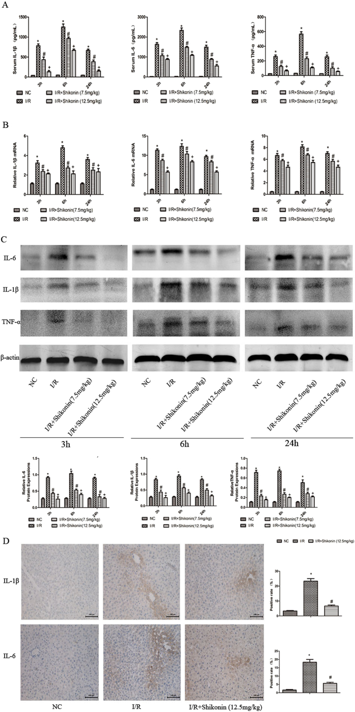
Hepatic ischemia/reperfusion (I/R) injury, which can result in severe liver injury and dysfunction, occurs in a variety of conditions such as liver transplantation, shock, and trauma. Cell death in hepatic I/R injury has been linked to apoptosis and autophagy. Shikonin plays a significant protective role in ischemia/reperfusion injury. The purpose of the present study was to investigate the protective effect of shikonin on hepatic I/R injury and explore the underlying mechanism. Mice were subjected to segmental (70%) hepatic warm ischemia to induce hepatic I/R injury. Two doses of shikonin (7.5 and 12.5 mg/kg) were administered 2 h before surgery. Balb/c mice were randomly divided into four groups: normal control, I/R, and shikonin preconditioning at two doses (7.5 and 12.5 mg/kg). The serum and liver tissues were collected at three time points (3, 6, and 24 h). Shikonin significantly reduced serum AST and ALT levels and improved pathological features. Shikonin affected the expression of Bcl-2, Bax, caspase 3, caspase 9, Beclin-1, and LC3, and upregulated PI3K and p-Akt compared with the levels in the I/R group. Shikonin attenuated hepatic I/R injury by inhibiting apoptosis and autophagy through a mechanism involving the activation of PI3K/Akt signaling.


Conflict of interest statement

The authors declare no competing financial interests.

Figures

Figure 1
Figure 1. Shikonin at two doses and 2% DMSO had no injurious effects on liver function.
(A) Serum AST and ALT levels were expressed as the mean ± SD (n = 6, P > 0.05). (B) The protein expression of Bcl-2, Bax, caspase 3, P62, Beclin-1, and LC3 was assessed by western blotting. The experiments were repeated three times. (C) Representative hematoxylin-and-eosin (HE) stained sections of liver tissues. Original magnification, ×200.
Figure 2
Figure 2. Pretreatment with shikonin ameliorated hepatic I/R injury.
(A) Serum AST and ALT levels were expressed as the mean ± SD (n = 6, *P < 0.05 for I/R versus NC, #P < 0.05 for I/R + Shikonin [7.5 mg/kg] versus I/R, +P < 0.05 for I/R + Shikonin [12.5 mg/kg] versus I/R). (B) The necrotic area stained with HE was analyzed with Image-Pro Plus 6.0 (magnification, ×200). The results showed statistically significant differences (n = 6, *P < 0.05 for I/R versus NC, #P < 0.05 for I/R + Shikonin [7.5 mg/kg] versus I/R, +P < 0.05 for I/R + Shikonin [12.5 mg/kg] versus I/R).
Figure 3
Figure 3. Shikonin reduced the release of IL-1β, TNF-α, and IL-6.
(A) The serum levels of IL-1β, TNF-α, and IL-6 are shown as the mean ± SD (n = 6, *P < 0.05 for I/R versus NC, #P < 0.05 for I/R + Shikonin [7.5 mg/kg] versus I/R, +P < 0.05 for I/R + Shikonin [12.5 mg/kg] versus I/R). (B) The mRNA expression of IL-1β, TNF-α, and IL-6 was assessed by RT-PCR. The experiments were repeated three times and the data are shown as the mean ± SD (n = 6, *P < 0.05 for I/R versus NC, #P < 0.05 for I/R + Shikonin [7.5 mg/kg] versus I/R, +P < 0.05 for I/R + Shikonin [12.5 mg/kg] versus I/R). (C) The protein expression of IL-1β, TNF-α, and IL-6 was determined by western blotting and the gray values were calculated (n = 6, *P < 0.05 for I/R versus NC, #P < 0.05 for I/R + Shikonin [7.5 mg/kg] versus I/R, +P < 0.05 for I/R + Shikonin [12.5 mg/kg] versus I/R). (D) Representative immunohistochemical staining (×200) showing the expression of IL-1β and IL-6 at 6 h. The ratio of brown areas to the total area was analyzed with Image-Pro Plus 6.0 (n = 6, *P < 0.05 for I/R versus NC, #P < 0.05 for I/R + Shikonin versus I/R).
Figure 4
Figure 4. Shikonin reduced apoptosis in hepatic I/R injury.
(A) The mRNA expression of Bcl-2, Bax, caspase 9, and caspase 3 was measured by RT-PCR and expressed as the mean ± SD (n = 6, *P < 0.05 for I/R versus NC, #P < 0.05 for I/R + Shikonin [7.5 mg/kg] versus I/R, +P < 0.05 for I/R + Shikonin [12.5 mg/kg] versus I/R). (B) The protein expression of Bcl-2, Bax, caspase 9, and caspase 3 was detected by western blotting and the gray values were calculated (n = 6, *P < 0.05 for I/R versus NC, #P < 0.05 for I/R + Shikonin [7.5 mg/kg] versus I/R, +P < 0.05 for I/R + Shikonin [12.5 mg/kg] versus I/R). (C) Immunohistochemical staining showing the expression of Bcl-2, Bax, and caspase 3 at 6 h. The ratio of brown areas to the total area was analyzed (n = 6, *P < 0.05 for I/R versus NC, #P < 0.05 for I/R + Shikonin versus I/R). (D) TUNEL staining (×200) of liver tissues at 6 h to detect apoptotic cell (n = 6, *P < 0.05 for I/R versus NC, #P < 0.05 for I/R + Shikonin versus I/R).
Figure 5
Figure 5. Shikonin inhibited autophagy in hepatic I/R injury.
(A) The mRNA expression of LC3, Beclin-1, and P62 was measured by RT-PCR and expressed as the mean ± SD (n = 6, *P < 0.05 for I/R versus NC, #P < 0.05 for I/R + Shikonin [7.5 mg/kg] versus I/R, +P < 0.05 for I/R + Shikonin [12.5 mg/kg] versus I/R). (B) The protein levels of LC3, Beclin-1, and P62 were detected by western blotting and the gray values were calculated (n = 6, *P < 0.05 for I/R versus NC, #P < 0.05 for I/R + Shikonin [7.5 mg/kg] versus I/R, +P < 0.05 for I/R + Shikonin [12.5 mg/kg] versus I/R). (C) Representative immunohistochemical staining (×200) showing the expression of LC3 and Beclin-1 at 6 h. The ratio of brown areas to the total area was analyzed with Image-Pro Plus (n = 6, *P < 0.05 for I/R versus NC, #P < 0.05 for I/R + Shikonin versus I/R). (D) Arrows indicate autophagosomes detected by transmission electron microscopy (TEM). Magnification: 20,000×.
Figure 6
Figure 6. Shikonin ameliorated hepatic I/R injury by activating the PI3K/Akt pathway.
(A) The mRNA expression of PI3K and Akt was detected by RT-PCR and the data are expressed as the mean ± SD (n = 6, *P < 0.05 for I/R versus NC, #P < 0.05 for I/R + Shikonin [7.5 mg/kg] versus I/R, +P < 0.05 for I/R + Shikonin [12.5 mg/kg] versus I/R). (B) The protein expression of PI3K, Akt, and p-Akt was detected by western blotting and the gray values were calculated (n = 6, *P < 0.05 for I/R versus NC, #P < 0.05 for I/R + Shikonin [7.5 mg/kg] versus I/R, +P < 0.05 for I/R + Shikonin [12.5 mg/kg] versus I/R). (C) Immunohistochemical staining (×200) showing the expression of PI3K and p-Akt at 6 h. The ratio of brown areas to the total area was analyzed with Image-Pro Plus (n = 6, *P < 0.05 for I/R versus NC, #P < 0.05 for I/R + Shikonin versus I/R).
Figure 7
Figure 7. Mechanism of the protective effects of shikonin on hepatic I/R injury.
Shikonin possesses anti-inflammatory effect and decreases the levels of pro-inflammatory cytokines such as IL-1β, IL-6, and TNF-α. Shikonin activates PI3K and promotes the phosphorylation of its downstream Akt, leading to the proliferation of injured hepatocytes. Phosphorylated Akt promotes the expression of Bcl-2, which inhibits the release of cytoC and down-regulates the expression of caspase 3 and caspase 9, leading to the inhibition of apoptosis. The upregulation of Bcl-2 inhibits the dissociation of Beclin1 from Bcl-2 and results in the reduction of Beclin-1, leading to the inhibition of autophagy.

Similar articles

Cited by

References

    1. Selzner N., Rudiger H., Graf R. & Clavien P. A. Protective strategies against ischemic injury of the liver. Gastroenterology 125, 917 (2003). - PubMed
    1. Gao L. et al.. Caveolin-1 protects against hepatic ischemia/reperfusion injury through ameliorating peroxynitrite-mediated cell death. Free Radic Biol Med 95, 209 (2016). - PubMed
    1. Karatzas T., Neri A. A., Baibaki M. E. & Dontas I. A. Rodent models of hepatic ischemia-reperfusion injury: time and percentage-related pathophysiological mechanisms. J Surg Res 191, 399 (2014). - PubMed
    1. Czubkowski P., Socha P. & Pawlowska J. Oxidative stress in liver transplant recipients. Ann Transplant 16, 99 (2011). - PubMed
    1. Siriussawakul A., Zaky A. & Lang J. D. Role of nitric oxide in hepatic ischemia-reperfusion injury. World J Gastroenterol 16, 6079 (2010). - PMC - PubMed

Publication types

MeSH terms