. 2017:3:25.
doi: 10.1051/sicotj/2017014. Epub 2017 Mar 22.

Lengthening and deformity correction about the knee using a magnetic internal lengthening nail

Affiliations

Lengthening and deformity correction about the knee using a magnetic internal lengthening nail

Austin T Fragomen et al. SICOT J. 2017.

Abstract

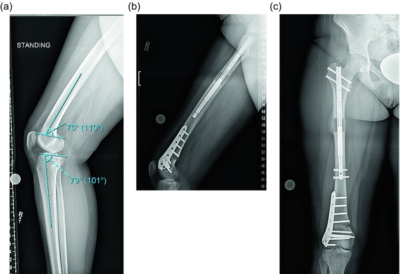
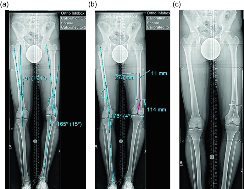
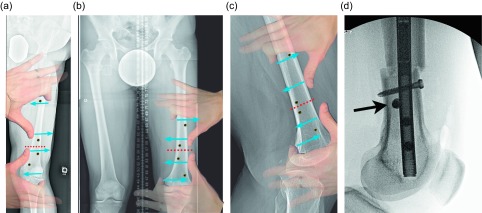
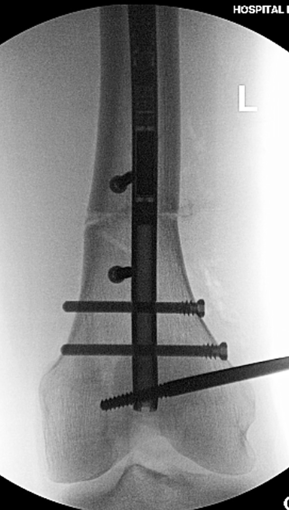
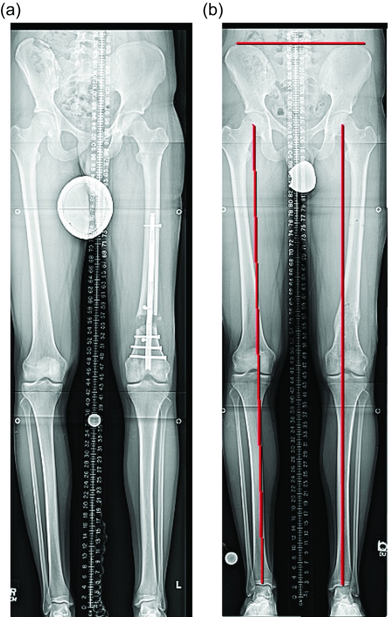
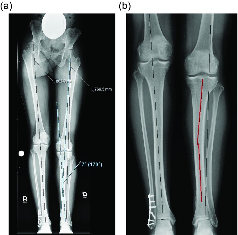
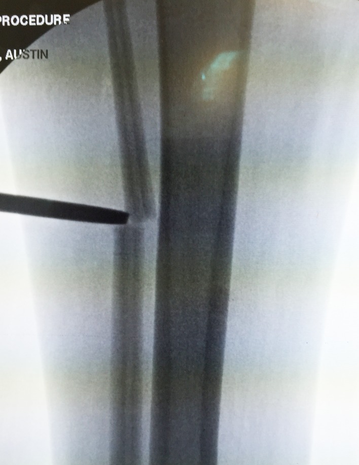
Introduction: The introduction of the internal lengthening nail (ILN) has changed the treatment of complex malalignment and shortening about the knee. Acute correction of the deformity and gradual lengthening through this osteotomy site has greatly simplified postoperative recovery. This manuscript is a review of the techniques that are currently being used in surgery.

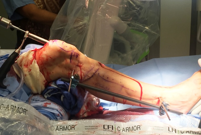
Methods: The article is broken into two sections: distal femur osteotomy and tibia osteotomy. Each is addressed separately since they have different personalities. Also included are topics of particular interest that surface in ongoing conferences regarding the ILN. This work is a mix of expert opinion and best practice supported by peer reviewed publications on the topic.

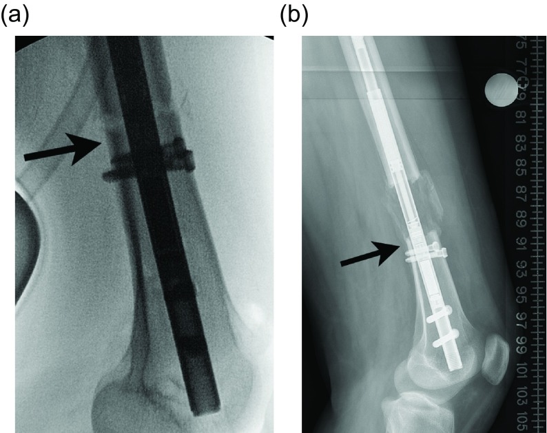
Results: Most published series demonstrate excellent results with the ILN. Certain precautions are reiterated including avoiding mechanical failure, need for a percutaneous osteotomy, need for over-reaming, and the need for blocking screws.

Discussion: Current controversies will be brought to light and discussed. The reader should find this aspect particularly helpful in navigating this rapidly evolving field.


Figures

Figure 1.
Figure 1.
(a) This preop lateral radiograph shows a 13° flexion deformity of the distal femur. (b) Postop lateral shows a correction of the apex anterior deformity through an posterior opening wedge osteotomy stabilized with a plate and a proximal femoral lengthening with an ILN. (c) This far distal osteotomy would be difficult to control with an intramedullary implant.
Figure 2.
Figure 2.
(a) This 51 inch AP radiograph allows for deformity planning with correction of varus and shortening. The mechanical axis planning is used to determine the magnitude and location of the deformity. (b) The red lines show the planned path of the ILN that will ensure correction of mechanical axis. The lines also show the length of the nail. (c) The white lines are placed to designate the path of the nail, and the circles are used to mark the location of the blocking screws. These radiographic plans are brought into the OR for comparison with intraop fluoroscopy shots.
Figure 3.
Figure 3.
(a) This illustration shows the suggested position of the blocking screws for correction of a valgus deformity. The screws centered around the osteotomy are more typically used leaving the peripheral screws as an option. (b) The same planning can be used for correction of a varus deformity. The overlay of the hands is showing a principle called the “reverse rule of thumbs” whereby the bone is grabbed with the thumb and index fingers of both hands and a correction simulated. The blocking screws should be placed opposite the location of the thumb and index fingers. (c) In the sagittal plane, the blocking screws are inserted posterior to the ILN at the osteotomy site. The distal screw is the most important, but the proximal screw can also be used for a shorter nail. The peripheral screws are seldom needed. (d). This fluoroscopy shot shows ideal posterior blocking screw placement: close to the osteotomy site and lying against the ILN (arrow).
Figure 4.
Figure 4.
A 6 mm half pin is seen in the typical location posterior to the path of the ILN.
Figure 5.
Figure 5.
A simple pin-to-bar frame is used to hold the reduction of the osteotomy.
Figure 6.
Figure 6.
The AP fluoroscopy shot shows the distal femur after successful distal interlocking with the varus deformity corrected. The peri-osteotomy blocking screws are positioned to prevent varus deviation during lengthening. The external half pin is also seen in the field.
Figure 7.
Figure 7.
(a) The lengthening is complete and the limb length is checked. The regenerate bone is filling in but is still plastic enough to allow for fine-tuning of the length. The long-standing X-ray can be affected by hip and knee joint muscle contractures yielding a false length measurement. This needs to be considered before over-lengthening the limb. (b) The final alignment and length are checked after ILN removal.
Figure 8.
Figure 8.
This intraop fluoroscopy frame shows a radio-opaque blush at the lateral aspect of the osteotomy site (arrow).
Figure 9.
Figure 9.
(a) The intraop fluoroscopy image shows contact of the posterior cortex of the distal fragment with the ILN at the osteotomy site (arrow). (b) After 5 cm of lengthening the distal fragment has not been able to flex due to the cortical abutment (arrow).
Figure 10.
Figure 10.
(a) The 51 inch standing film shows a diaphyseal valgus deformity and limb length discrepancy. (b) The path of the ILN is planned with the osteotomy site at the intersection of the red lines.
Figure 11.
Figure 11.
(a) This shows a different patient’s X-rays. The valgus correction is maintained by placing blocking screws in the concavity of the deformity (lateral aspect). Peripheral blocking screws can be used as well. (b) The sagittal plane requires at least one blocking screw in the proximal fragment near the osteotomy (the screw shown at the entry site of the ILN is theoretical and is never used). (c) This lateral film shows a well-placed posterior blocking screw (arrow).
Figure 12.
Figure 12.
In this unrelated case, the external fixator helps to maintain the reduction of the deformity correction after osteotomy during reaming. A suprapatellar approach was used in this case.
Figure 13.
Figure 13.
The osteotome is inserted through a limited incision under fluoroscopy.
Figure 14.
Figure 14.
In this unrelated case, the proximal syndesmotic screw also served as the posterior blocking screw (arrow).
Figure 15.
Figure 15.
(a) The X-rays shown in Figures 15a and 15b are a continuation of the patient presented in Figures 10a and 10b. The end of distraction long-standing radiograph shows equal limb length and ideal alignment. (b) A lateral radiograph shows interval healing of the regenerate and ideal sagittal alignment.

References

    1. Landge V, Shabtai L, Gesheff Specht SC, Herzenberg JE (2015) Patient satisfaction after limb lengthening with internal and external devices. J Surg Orthop Adv 24(3), 174–179. - PubMed
    1. Karakoyun O, Kucukkaya M, Erol MF (2015) Does lengthening after acute correction negatively affect bone healing during distraction osteogenesis? Acta Orthop Traumatol Turc 49(4), 405–409. - PubMed
    1. Horn J, Grimsrud O, Dagsgard A, et al. (2015) Femoral lengthening with a motorized intramedullary nail. Acta Orthop 86(2), 248–256. - PMC - PubMed
    1. Burghardt RD, Paley D, Specht SC, Herzenberg JE (2012) The effect on mechanical axis deviation of femoral lengthening with an intramedullary telescopic nail. J Bone Joint Surg Br 94(9), 1241–1245. - PubMed
    1. Baumgart R (2009) The reverse planning method for lengthening of the lower limb using a straight intramedullary nail with or without deformity correction. A new method. Oper Orthop Traumatol 21(2), 221–233. - PubMed

LinkOut - more resources