Review
. 2017 May:86:37-41.
doi: 10.1016/j.biocel.2017.03.009. Epub 2017 Mar 18.

Platinum replica electron microscopy: Imaging the cytoskeleton globally and locally

Affiliations
Review

Platinum replica electron microscopy: Imaging the cytoskeleton globally and locally

Tatyana M Svitkina. Int J Biochem Cell Biol. 2017 May.

Abstract

Structural studies reveal how smaller components of a system work together as a whole. However, combining high resolution of details with full coverage of the whole is challenging. In cell biology, light microscopy can image many cells in their entirety, but at a lower resolution, whereas electron microscopy affords very high resolution, but usually at the expense of the sample size and coverage. Structural analyses of the cytoskeleton are especially demanding, because cytoskeletal networks are unresolvable by light microscopy due to their density and intricacy, whereas their proper preservation is a challenge for electron microscopy. Platinum replica electron microscopy can uniquely bridge the gap between the "comfort zones" of light and electron microscopy by allowing high resolution imaging of the cytoskeleton throughout the entire cell and in many cells in the population. This review describes the principles and applications of platinum replica electron microscopy for studies of the cytoskeleton.

Keywords: Actin filaments; Cytoskeleton; Electron microscopy; Platinum replica; Tomography.


Figures

Figure 1
Figure 1
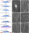
PREM of a human umbilical vein endothelial cell stained with anti-VE-cadherin immunogold. (A) Workflow of PREM of cultured cells. (B) Overview of the cell with labeled regions enlarged in panels C-G. (C, D) Actin filament networks in the lamellum (C) and the lamellipodium (D). (E) Enlarged region of the branched actin network from the box in D. (F) Junction between two cells. (G) Enlarged focal adherens junction from the box in F; gold particles labeling VE-cadherin are pseudocolored in yellow.
Figure 2
Figure 2
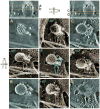
Electron tomography of an unroofed Ptk2 cell shows a clathrin-coated pit with the associated branched actin filament network (pseudocolored green in the central panel) abutting the constricted neck of the pit. Brown panels: dual-axis tilt images at the indicated angles. Blue panels: vertical section through the pit (top) and horizontal slices (A-D) along yellow lines with corresponding letters. Neck size and degree of invagination can be seen in D and in upper panels.

References

    1. Collins A, Warrington A, Taylor KA, Svitkina T. Structural organization of the actin cytoskeleton at sites of clathrin-mediated endocytosis. Curr Biol. 2011;21:1167–1175. - PMC - PubMed
    1. Heuser J. Protocol for 3-D visualization of molecules on mica via the quick-freeze, deep-etch technique. J Electron Microsc Tech. 1989;13:244–263. - PubMed
    1. Heuser J. The production of ‘cell cortices’ for light and electron microscopy. Traffic. 2000;1:545–552. - PubMed
    1. Heuser JE, Kirschner MW. Filament organization revealed in platinum replicas of freeze-dried cytoskeletons. J Cell Biol. 1980;86:212–234. - PMC - PubMed
    1. Hirokawa N. Cross-linker system between neurofilaments, microtubules, and membranous organelles in frog axons revealed by the quick-freeze, deep-etching method. J Cell Biol. 1982;94:129–142. - PMC - PubMed

Publication types

LinkOut - more resources