. 2017 Mar 21;13(3):e1006261.
doi: 10.1371/journal.ppat.1006261. eCollection 2017 Mar.

Ubiquitously expressed Human Beta Defensin 1 (hBD1) forms bacteria-entrapping nets in a redox dependent mode of action

Affiliations

Ubiquitously expressed Human Beta Defensin 1 (hBD1) forms bacteria-entrapping nets in a redox dependent mode of action

Judith Raschig et al. PLoS Pathog. .

Abstract

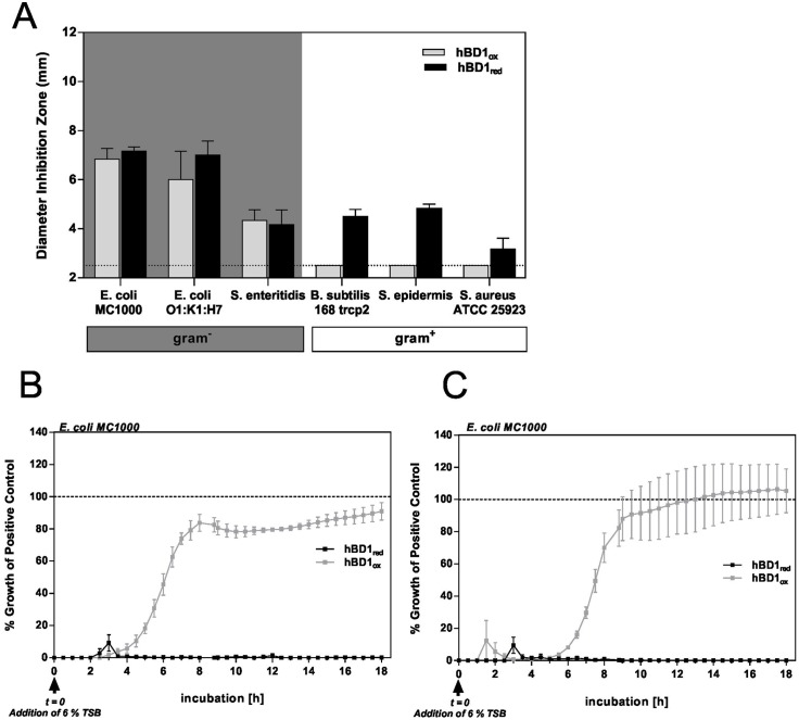
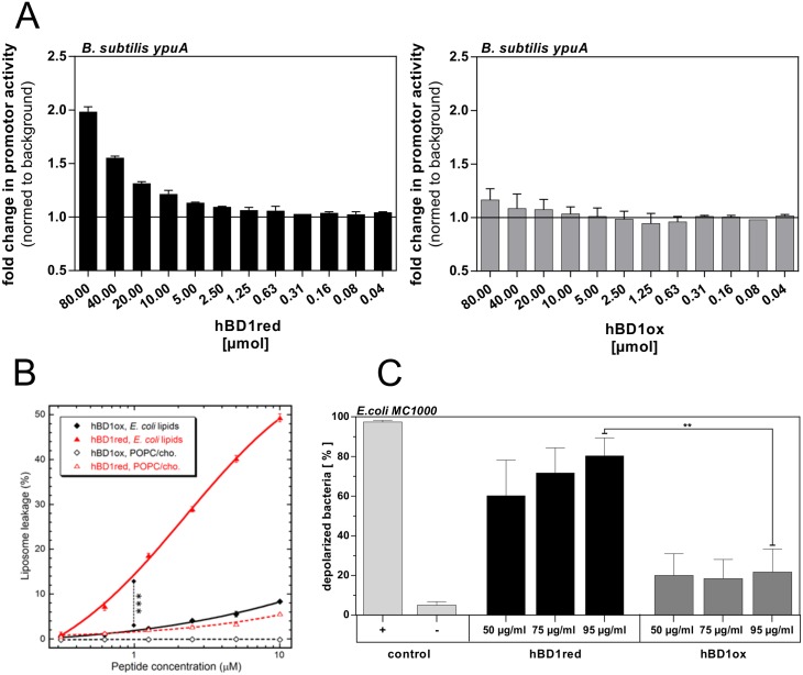
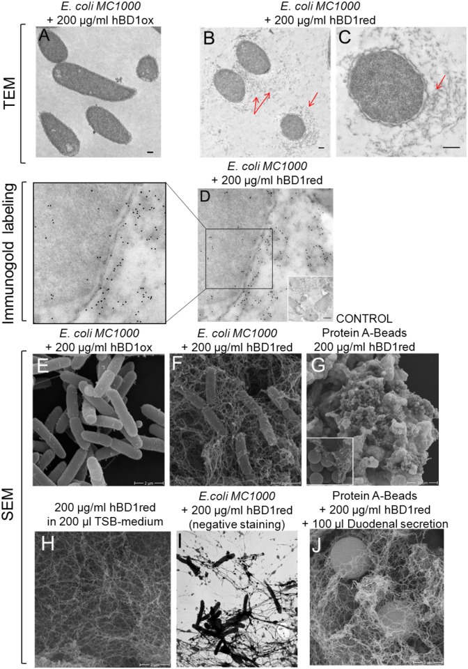
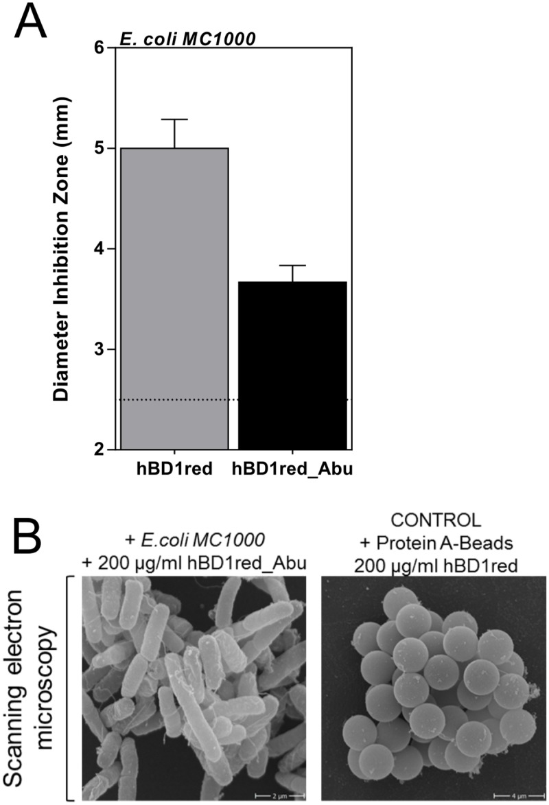
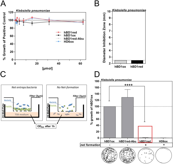
Ever since the discovery of endogenous host defense antimicrobial peptides it has been discussed how these evolutionary conserved molecules avoid to induce resistance and to remain effective. Human ß-defensin 1 (hBD1) is an ubiquitously expressed endogenous antimicrobial peptide that exhibits qualitatively distinct activities between its oxidized and reduced forms. Here, we explore these antimicrobial mechanisms. Surprisingly, using electron microscopy we detected a so far unknown net-like structure surrounding bacteria, which were treated with the reduced but not the oxidized form of hBD1. A transmigration assay demonstrated that hBD1-derived nets capture bacteria and inhibit bacterial transmigration independent of bacterial killing. The presence of nets could completely prevent migration of hBD1 resistant pathogens and are stable in the presence of human duodenal secretion with a high amount of proteases. In contrast to HD6, cysteins are necessary for net formation. This redox-dependent function serves as an additional mechanism of action for hBD1 and differs from net formation by other defensins such as Paneth cell-derived human α-defensin 6 (HD6). While hBD1red and hBD1ox have distinct antimicrobial profiles and functions, only the reduced form provides additional host protection by entrapping bacteria in extracellular net structures preventing bacterial invasion. Better understanding of the modes of action of endogenous host peptides will help to find new antimicrobial strategies.


Conflict of interest statement

The authors have declared that no competing interests exist.

Figures

Fig 1
Fig 1. Antimicrobial activity of hBD1 is dependent of its redox status.
Bacteria were incubated with hBD1ox and hBD1red in (A) Radial Diffusion Assay. Antimicrobial activity was measured by analyzing the diameter of the inhibition zone. A diameter of 2.5 mm (dotted line) is the diameter of the punched well (n = 3). As it is not possible to distinguish between a bactericidal and a bacteriostatic mechanism using RDA as an end-point determination, we additionally performed a time-course in a turbidity broth assay. Bacteria were pretreated with 62.5 μM (B) and 125 μM (C) of the peptides before addition of 6% TSB and % growth of the untreated control over the following 18 h was plotted. In contrast to hBD1red, the E. coli culture treated with hBD1ox is able to regrow with a delay of 4 h (B) and 5.5 h (C) compared to the control. Data are presented as mean +/- SEM of at least two (B+C) or three (A) independent experiments.
Fig 2
Fig 2. Characterization of the hBD1red target in bacterial compartments.
(A) Transcription activity of the B. subtilis ypuA promoter indicative of cell envelope damage. Bacteria were incubated with increasing concentrations of hBD1ox and hBD1red. (B) Membrane leakage as a result of hBD1 treatment was determined by quantifying carboxyfluorescein efflux from liposomes POPC/cholesterol (3:2 molar ratio, a human membrane model) or an E. coli polar lipid extract. Presented is the leakage level reached after 45 min incubation with mean and standard deviations from triplicates. Permeabilization, by hBD1red on E. coli liposomes, was significantly higher than by hBD1ox (***p<0.001 only for 1 μM). (C) Membrane depolarization of 1.5 x 10⁶ CFU E. coli MC1000 in response to hBD1. 1 h treatment was analyzed by flow cytometry. Positive control (+) was incubated with hBD3ox (50 μg/ml) and the negative control (-) without any peptide. The statistic was evaluated by using student’s t-test with **p = 0.0072. Data are presented as mean +/- SEM of at least three independent experiments.
Fig 3
Fig 3. HBD1red forms netlike structures.
Bacteria or beads were treated with hBD1 redox forms for 2 h. Samples were fixed and visualized by electron microscopy. (A-C) Transmission electron microscopy of E. coli MC1000 for analyzing the bacterial morphology. HBD1red-treated E. coli cells display strong membrane defects and an unknown grey structure around the bacteria was detected. (D) E. coli MC1000 was incubated with hBD1red and with antibodies against the peptide. The secondary antibodies are conjugated to gold particles, 6 nm (black points). (E) E. coli MC1000 were treated with hBD1ox. (F-I) Bacteria or protein A-coated beads were treated with hBD1red and investigated by scanning electron microscopy. (G) As a control we used hBD1red with beads (H) and alone in medium without bacteria and beads. (I) HBD1red net formation in native samples without addition of fixative agents. (J) Protein A-coated beads were incubated with hBD1red and human duodenal secretion, which was taken from one individual.
Fig 4
Fig 4. Net-formation of hBD1red depends on free cysteines.
(A) 2 μg of hBD1red and hBD1red_Abu, a variant in which cysteine is replaced by α-amino-butyric acid (Abu), were tested in a RDA Assay against E. coli MC1000 or (B) 200 μg/ ml peptide with bacteria or protein A-coated beads were analyzed by scanning electron microscopy. Data in A) are presented as mean +/- SEM of at least three independent experiments.
Fig 5
Fig 5. Proof for functional net-formation of hBD1red.
Comparison of antimicrobial activity of different hBD1 forms. Antimicrobial activity of hBD1 forms was compared in a (A) turbidity broth assay or (B) RDA against K. pneumoniae. Different concentrations of peptides were incubated with bacteria and change in optical density (OD600) was measured after 12 h or the diameter of inhibition zone was analyzed. HBD1 redox forms are not antimicrobial active against K. pneumoniae. (C) Schematic model for the Transwell Membrane Assay. Bacteria are entrapped in a net and are not able to pass the porous membrane (left), but in absence of nets bacteria pass through the membrane and grow in the TSB medium in the chamber below. (D) Bacterial growth was measured at OD600 after 1 h pre-incubation of peptide in filter and additional 2 h and % growth of negative control was plotted. With net formation no significant numbers of bacteria are able to translocate which is in contrast to hBD1ox or hBD1red_Abu with no net formation. Representative blood agar plates are shown. Data are presented as mean +/- SEM of at least three independent experiments. The statistic was evaluated by using student’s t-test with ****p < 0.0001.

References

    1. Ostaff MJ, Stange EF, Wehkamp J. Antimicrobial peptides and gut microbiota in homeostasis and pathology. EMBO Mol Med. 2013;5: 1–19. - PMC - PubMed
    1. Diamond G, Beckloff N, Weinberg A, Kisich KO. The roles of antimicrobial peptides in innate host defense. Curr Pharm Des. 2009;15: 2377–2392. - PMC - PubMed
    1. Zasloff M. Antimicrobial peptides of multicellular organisms. Nature. 2002;415: 389–395. 10.1038/415389a - DOI - PubMed
    1. Bevins CL. Antimicrobial peptides as effector molecules of mammalian host defense. Contrib Microbiol. 2003;10: 106–148. - PubMed
    1. Nizet V, Cole JN. Bacterial Evasion of Host Antimicrobial Peptide Defenses. Microbiol Spectr. 2016;4. - PMC - PubMed

Publication types

LinkOut - more resources